
The passage of a sound wave in the human hearing organ. Structure and functions of the outer, middle and inner ear
Peripheral part auditory analyzer In humans, it is morphologically united with the peripheral part of the vestibular analyzer, and morphologists call this structure organelukha and balance (organum vestibulo-cochleare). It has three sections:
- outer ear (external ear canal, auricle with muscles and ligaments);
- middle ear (tympanic cavity, mastoid appendages, auditory tube)
- inner ear(membranous labyrinth located in the bony labyrinth inside the pyramid temporal bone).
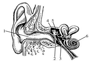
1. The outer ear concentrates sound vibrations and directs them to the external auditory opening.
2. The auditory canal conducts sound vibrations to the eardrum
3. The eardrum is a membrane that vibrates when exposed to sound.
4. The hammer is attached to the center with its handle eardrum with the help of ligaments, and its head is connected to the incus (5), which, in turn, is attached to the stapes (6).
Tiny muscles help transmit sound by regulating the movement of these ossicles.
7. The Eustachian (or auditory) tube connects the middle ear to the nasopharynx. When the ambient air pressure changes, the pressure on both sides of the eardrum is equalized through the auditory tube.
8. Vestibular system. The vestibular system in our ear is part of the body's balance system. Sensory cells provide information about the position and movement of our head.
9. The cochlea is the organ of hearing directly connected to the auditory nerve. The name of the snail is determined by its spirally convoluted shape. This is a bone canal that forms two and a half turns of a spiral and is filled with fluid. The anatomy of the cochlea is very complex, and some of its functions are still unexplored.
The organ of Corti consists of a number of sensory, hair-bearing cells (12) that cover the basilar membrane (13). Sound waves are picked up by hair cells and converted into electrical impulses. These electrical impulses are then transmitted along the auditory nerve (11) to the brain. The auditory nerve consists of thousands of tiny nerve fibers. Each fiber starts from a specific part of the cochlea and transmits a specific sound frequency. Low-frequency sounds are transmitted through fibers emanating from the apex of the cochlea (14), and high-frequency sounds are transmitted through fibers connected to its base. Thus, the function of the inner ear is to convert mechanical vibrations into electrical ones, since the brain can only perceive electrical signals.
Outer ear is a sound-collecting device. The external auditory canal conducts sound vibrations to the eardrum. The eardrum, which separates the outer ear from the tympanic cavity, or middle ear, is a thin (0.1 mm) partition shaped like an inward funnel. The membrane vibrates with action sound vibrations, coming to her through the external auditory canal.
Sound vibrations are picked up by the ears (in animals they can turn towards the sound source) and transmitted through the external auditory canal to the eardrum, which separates the outer ear from the middle ear. Catching sound and the entire process of listening with two ears - so-called binaural hearing - is important for determining the direction of sound. Sound vibrations coming from the side reach the nearest ear a few ten-thousandths of a second (0.0006 s) earlier than the other. This insignificant difference in the time of arrival of sound to both ears is enough to determine its direction.
Middle ear is a sound-conducting device. It is an air cavity that connects to the nasopharynx cavity through the auditory (Eustachian) tube. Vibrations from the eardrum through the middle ear are transmitted by 3 auditory ossicles connected to each other - the hammer, incus and stapes, and the latter through the membrane oval window transmits these vibrations to the fluid located in the inner ear - perilymph.
Due to the geometric features auditory ossicles vibrations of the tympanic membrane of reduced amplitude but increased strength are transmitted to the stapes. In addition, the surface of the stapes is 22 times smaller than the eardrum, which increases its pressure on the oval window membrane by the same amount. As a result of this, even weak sound waves acting on the eardrum can overcome the resistance of the membrane of the oval window of the vestibule and lead to vibrations of the fluid in the cochlea.
With strong sounds, special muscles reduce the mobility of the eardrum and auditory ossicles, adapting hearing aid to such changes in the stimulus and protecting the inner ear from destruction.
Thanks to the connection through the auditory tube of the air cavity of the middle ear with the cavity of the nasopharynx, it becomes possible to equalize the pressure on both sides of the eardrum, which prevents its rupture during significant changes in pressure during external environment- when diving under water, climbing to heights, shooting, etc. This is the barofunction of the ear.
There are two muscles in the middle ear: the tensor tympani and the stapedius. The first of them, contracting, increases the tension of the eardrum and thereby limits the amplitude of its vibrations during strong sounds, and the second fixes the stapes and thereby limits its movements. The reflex contraction of these muscles occurs 10 ms after the onset of a strong sound and depends on its amplitude. This automatically protects the inner ear from overload. For instantaneous strong irritations (impacts, explosions, etc.) this defense mechanism does not have time to work, which can lead to hearing impairment (for example, among bombers and artillerymen).
Inner ear is a sound-perceiving apparatus. It is located in the pyramid of the temporal bone and contains the cochlea, which in humans forms 2.5 spiral turns. The cochlear canal is divided by two partitions, the main membrane and the vestibular membrane into 3 narrow passages: upper (scala vestibular), middle (membranous canal) and lower (scala tympani). At the top of the cochlea there is an opening that connects the upper and lower canals into a single one, going from the oval window to the top of the cochlea and then to the round window. Its cavity is filled with fluid - peri-lymph, and the cavity of the middle membranous canal is filled with a fluid of a different composition - endolymph. In the middle channel there is a sound-perceiving apparatus - the organ of Corti, in which there are mechanoreceptors of sound vibrations - hair cells.
The main route of delivery of sounds to the ear is airborne. The approaching sound vibrates the eardrum, and then through the chain of auditory ossicles the vibrations are transmitted to the oval window. At the same time, vibrations of the air in the tympanic cavity also occur, which are transmitted to the membrane of the round window. Another way of delivering sounds to the cochlea is fabric or bone conduction . In this case, the sound directly acts on the surface of the skull, causing it to vibrate. Bone pathway for sound transmission becomes of great importance if a vibrating object (for example, the stem of a tuning fork) comes into contact with the skull, as well as in diseases of the middle ear system, when the transmission of sounds through the chain of auditory ossicles is disrupted. Except air route, the conduction of sound waves exists through a tissue, or bone, path. Under the influence of air sound vibrations, as well as when vibrators (for example, a bone telephone or a bone tuning fork) come into contact with the integument of the head, the bones of the skull begin to vibrate (the bone labyrinth also begins to vibrate). Based on the latest data (Bekesy and others), it can be assumed that sounds propagating along the bones of the skull only excite the organ of Corti if, similar to air waves, they cause arching of a certain section of the main membrane. The ability of the skull bones to conduct sound explains why to the person himself his voice, recorded on tape, seems alien when the recording is played back, while others easily recognize it. The fact is that the tape recording does not reproduce your entire voice. Usually, when talking, you hear not only those sounds that your interlocutors also hear (that is, those sounds that are perceived due to air-liquid conduction), but also those low-frequency sounds, the conductor of which is the bones of your skull. However, when listening to a tape recording of your own voice, you hear only what could be recorded - sounds whose conductor is air. Binaural hearing . Humans and animals have spatial hearing, that is, the ability to determine the position of a sound source in space. This property is based on the presence of binaural hearing, or listening with two ears. It is also important for him to have two symmetrical halves at all levels of the auditory system. The acuity of binaural hearing in humans is very high: the position of the sound source is determined with an accuracy of 1 angular degree. The basis for this is the ability of the neurons of the auditory system to evaluate interaural (interaural) differences in the time of sound arrival on the right and left ear and sound intensity in each ear. If the sound source is located away from the midline of the head, the sound wave arrives at one ear slightly earlier and has greater strength than at the other ear. Assessing the distance of a sound source from the body is associated with a weakening of the sound and a change in its timbre.When the right and left ears are stimulated separately via headphones, a delay between sounds of as little as 11 µs or a 1 dB difference in the intensity of the two sounds results in an apparent shift in the localization of the sound source from the midline towards an earlier or stronger sound. The auditory centers contain neurons that are acutely tuned to a specific range of interaural differences in time and intensity. Cells have also been found that respond only to a certain direction of movement of a sound source in space.
Information . Physiology of VNI and sensory systems . Fundamentals of neurophysiology and GNI .
The peripheral part of the auditory analyzer is morphologically combined in humans with the peripheral part of the vestibular analyzer, and morphologists call this structure the organum vestibulo-cochleare. It has three sections:
· external ear (external auditory canal, auricle with muscles and ligaments);
middle ear (tympanic cavity, mastoid appendages, auditory tube)
· inner ear (membranous labyrinth located in the bony labyrinth inside the pyramid of the temporal bone).
External ear (external auditory canal, pinna with muscles and ligaments)
Middle ear (tympanic cavity, mastoid appendages, auditory tube)
Inner ear (membranous labyrinth located in the bony labyrinth inside the pyramid of the temporal bone)
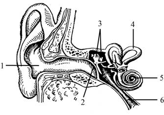
1. The outer ear concentrates sound vibrations and directs them to the external auditory opening.
2. The auditory canal conducts sound vibrations to the eardrum
3. The eardrum is a membrane that vibrates when exposed to sound.
4. The malleus with its handle is attached to the center of the eardrum with the help of ligaments, and its head is connected to the incus (5), which, in turn, is attached to the stapes (6).
Tiny muscles help transmit sound by regulating the movement of these ossicles.
7. The Eustachian (or auditory) tube connects the middle ear to the nasopharynx. When the ambient air pressure changes, the pressure on both sides of the eardrum is equalized through the auditory tube.
8. Vestibular system. The vestibular system in our ear is part of the body's balance system. Sensory cells provide information about the position and movement of our head.
9. The cochlea is the organ of hearing directly connected to the auditory nerve. The name of the snail is determined by its spirally convoluted shape. This is a bone canal that forms two and a half turns of a spiral and is filled with fluid. The anatomy of the cochlea is very complex, and some of its functions are still unexplored.
Organ of Corti
The organ of Corti consists of a number of sensory, hair-bearing cells (12) that cover the basilar membrane (13). Sound waves are picked up by hair cells and converted into electrical impulses. These electrical impulses are then transmitted along the auditory nerve (11) to the brain. The auditory nerve consists of thousands of tiny nerve fibers. Each fiber starts from a specific part of the cochlea and transmits a specific sound frequency. Low-frequency sounds are transmitted through fibers emanating from the apex of the cochlea (14), and high-frequency sounds are transmitted through fibers connected to its base. Thus, the function of the inner ear is to convert mechanical vibrations into electrical ones, since the brain can only perceive electrical signals.
Outer ear is a sound-collecting device. The external auditory canal conducts sound vibrations to the eardrum. The eardrum, which separates the outer ear from the tympanic cavity, or middle ear, is a thin (0.1 mm) partition shaped like an inward funnel. The membrane vibrates under the action of sound vibrations coming to it through the external auditory canal.
Sound vibrations are picked up by the ears (in animals they can turn towards the sound source) and transmitted through the external auditory canal to the eardrum, which separates the outer ear from the middle ear. Catching sound and the entire process of listening with two ears - so-called binaural hearing - is important for determining the direction of sound. Sound vibrations coming from the side reach the nearest ear a few ten-thousandths of a second (0.0006 s) earlier than the other. This insignificant difference in the time of arrival of sound to both ears is enough to determine its direction.
Middle ear is a sound-conducting device. It is an air cavity that connects to the nasopharynx cavity through the auditory (Eustachian) tube. Vibrations from the eardrum through the middle ear are transmitted by 3 auditory ossicles connected to each other - the hammer, incus and stapes, and the latter, through the membrane of the oval window, transmits these vibrations to the fluid located in the inner ear - perilymph.
Due to the peculiarities of the geometry of the auditory ossicles, vibrations of the eardrum of reduced amplitude but increased strength are transmitted to the stapes. In addition, the surface of the stapes is 22 times smaller than the eardrum, which increases its pressure on the oval window membrane by the same amount. As a result of this, even weak sound waves acting on the eardrum can overcome the resistance of the membrane of the oval window of the vestibule and lead to vibrations of the fluid in the cochlea.
During strong sounds, special muscles reduce the mobility of the eardrum and auditory ossicles, adapting the hearing aid to such changes in the stimulus and protecting the inner ear from destruction.
Thanks to the connection of the air cavity of the middle ear with the cavity of the nasopharynx through the auditory tube, it becomes possible to equalize the pressure on both sides of the eardrum, which prevents its rupture during significant changes in pressure in the external environment - when diving under water, climbing to a height, shooting, etc. This is the barofunction of the ear .
There are two muscles in the middle ear: the tensor tympani and the stapedius. The first of them, contracting, increases the tension of the eardrum and thereby limits the amplitude of its vibrations during strong sounds, and the second fixes the stapes and thereby limits its movements. The reflex contraction of these muscles occurs 10 ms after the onset of a strong sound and depends on its amplitude. This automatically protects the inner ear from overload. In case of instantaneous strong irritations (impacts, explosions, etc.), this protective mechanism does not have time to work, which can lead to hearing impairment (for example, among bombers and artillerymen).
Inner ear is a sound-perceiving apparatus. It is located in the pyramid of the temporal bone and contains the cochlea, which in humans forms 2.5 spiral turns. The cochlear canal is divided by two partitions, the main membrane and the vestibular membrane into 3 narrow passages: upper (scala vestibular), middle (membranous canal) and lower (scala tympani). At the top of the cochlea there is an opening that connects the upper and lower canals into a single one, going from the oval window to the top of the cochlea and then to the round window. Its cavity is filled with fluid - peri-lymph, and the cavity of the middle membranous canal is filled with a fluid of a different composition - endolymph. In the middle channel there is a sound-perceiving apparatus - the organ of Corti, in which there are mechanoreceptors of sound vibrations - hair cells.
The main route of delivery of sounds to the ear is airborne. The approaching sound vibrates the eardrum, and then through the chain of auditory ossicles the vibrations are transmitted to the oval window. At the same time, vibrations of the air in the tympanic cavity also occur, which are transmitted to the membrane of the round window.
Another way of delivering sounds to the cochlea is tissue or bone conduction . In this case, the sound directly acts on the surface of the skull, causing it to vibrate. Bone pathway for sound transmission becomes of great importance if a vibrating object (for example, the stem of a tuning fork) comes into contact with the skull, as well as in diseases of the middle ear system, when the transmission of sounds through the chain of auditory ossicles is disrupted. In addition to the air path for conducting sound waves, there is a tissue, or bone, path.
Under the influence of airborne sound vibrations, as well as when vibrators (for example, a bone telephone or a bone tuning fork) come into contact with the integument of the head, the bones of the skull begin to vibrate (the bone labyrinth also begins to vibrate). Based on the latest data (Bekesy and others), it can be assumed that sounds propagating along the bones of the skull only excite the organ of Corti if, similar to air waves, they cause arching of a certain section of the main membrane.
The ability of the skull bones to conduct sound explains why to the person himself his voice, recorded on tape, seems alien when the recording is played back, while others easily recognize it. The fact is that the tape recording does not reproduce your entire voice. Usually, when talking, you hear not only those sounds that your interlocutors also hear (that is, those sounds that are perceived due to air-liquid conduction), but also those low-frequency sounds, the conductor of which is the bones of your skull. However, when listening to a tape recording of your own voice, you hear only what could be recorded - sounds whose conductor is air.
Binaural hearing. Humans and animals have spatial hearing, that is, the ability to determine the position of a sound source in space. This property is based on the presence of binaural hearing, or listening with two ears. It is also important for him to have two symmetrical halves at all levels of the auditory system. The acuity of binaural hearing in humans is very high: the position of the sound source is determined with an accuracy of 1 angular degree. The basis for this is the ability of neurons in the auditory system to evaluate interaural (inter-ear) differences in the time of arrival of sound to the right and left ear and the intensity of sound in each ear. If the sound source is located away from the midline of the head, the sound wave arrives at one ear slightly earlier and has greater strength than at the other ear. Assessing the distance of a sound source from the body is associated with a weakening of the sound and a change in its timbre.
When the right and left ears are stimulated separately via headphones, a delay between sounds of as little as 11 µs or a 1 dB difference in the intensity of the two sounds results in an apparent shift in the localization of the sound source from the midline towards an earlier or stronger sound. The auditory centers contain neurons that are acutely tuned to a specific range of interaural differences in time and intensity. Cells have also been found that respond only to a certain direction of movement of a sound source in space.
Consists of the outer, middle and inner ear. The middle and inner ear are located inside the temporal bone.
Outer ear consists of the auricle (collects sounds) and the external auditory canal, which ends in the eardrum.
Middle ear- This is a chamber filled with air. It contains the auditory ossicles (hammer, incus and stapes), which transmit vibrations from the eardrum to the membrane of the oval window - they amplify the vibrations 50 times. The middle ear is connected to the nasopharynx via the Eustachian tube, through which the pressure in the middle ear is equalized with atmospheric pressure.
In the inner ear there is a cochlea - a fluid-filled bone canal twisted in 2.5 turns, blocked by a longitudinal septum. On the septum there is an organ of Corti containing hair cells - these are auditory receptors that convert sound vibrations into nerve impulses.
Ear work: When the stapes presses on the membrane of the oval window, the column of fluid in the cochlea moves, and the membrane of the round window protrudes into the middle ear. The movement of the fluid causes the hairs to touch the integumentary plate, causing the hair cells to become excited.
Vestibular apparatus: in the inner ear, in addition to the cochlea, there are semicircular canals and vestibular sacs. Hair cells in the semicircular canals sense fluid movement and respond to acceleration; hair cells in the sacs sense the movement of the otolith pebble attached to them and determine the position of the head in space.
Establish a correspondence between the structures of the ear and the sections in which they are located: 1) outer ear, 2) middle ear, 3) inner ear. Write the numbers 1, 2 and 3 in the correct order.
A) auricle
B) oval window
B) snail
D) stirrup
D) Eustachian tube
E) hammer
Answer
Establish a correspondence between the function of the hearing organ and the section that performs this function: 1) middle ear, 2) inner ear
A) conversion of sound vibrations into electrical ones
B) amplification of sound waves due to vibrations of the auditory ossicles
B) equalization of pressure on the eardrum
D) conducting sound vibrations due to the movement of liquid
D) irritation of auditory receptors
Answer
1. Set the transmission sequence sound wave to auditory receptors. Write down the corresponding sequence of numbers.
1) vibrations of the auditory ossicles
2) vibrations of fluid in the cochlea
3) vibrations of the eardrum
4) irritation of auditory receptors
Answer
2. Install correct sequence passage of a sound wave through the human hearing organ. Write down the corresponding sequence of numbers.
1) eardrum
2) oval window
3) stirrup
4) anvil
5) hammer
6) hair cells
Answer
3. Establish the sequence in which sound vibrations are transmitted to the receptors of the hearing organ. Write down the corresponding sequence of numbers.
1) Outer ear
2) Oval window membrane
3) Auditory ossicles
4) Eardrum
5) Fluid in the cochlea
6) Hearing receptors
Answer
4. Establish the sequence of arrangement of the structures of the human ear, starting with the one that captures the sound wave. Write down the corresponding sequence of numbers.
1) oval window of the cochlea of the inner ear
2) external auditory canal
3) eardrum
4) auricle
5) auditory ossicles
6) organ of Corti
Answer
5. Establish the sequence of transmission of sound vibrations to the receptors of the human hearing organ. Write down the corresponding sequence of numbers.
1) external auditory canal
2) oval window membrane
3) auditory ossicles
4) eardrum
5) fluid in the cochlea
6) hair cells of the cochlea
Answer
1. Select three correctly labeled captions for the drawing “Structure of the Ear.”
1) external auditory canal
2) eardrum
3) auditory nerve
4) stirrup
5) semicircular canal
6) snail
Answer
2. Select three correctly labeled captions for the drawing “Structure of the Ear.” Write down the numbers under which they are indicated.
1) ear canal
2) eardrum
3) auditory ossicles
4) auditory tube
5) semicircular canals
6) auditory nerve
Answer

4. Select three correctly labeled captions for the drawing “Structure of the Ear.”
1) auditory ossicles
2) facial nerve
3) eardrum
4) auricle
5) middle ear
6) vestibular apparatus
Answer
1. Set the sequence of sound transmission in the hearing analyzer. Write down the corresponding sequence of numbers.
1) vibration of the auditory ossicles
2) vibration of fluid in the cochlea
3) generation nerve impulse
5) transmission of nerve impulses along the auditory nerve to the temporal lobe of the cortex cerebral hemispheres
6) vibration of the oval window membrane
7) vibration of hair cells
Answer
2. Establish the sequence of processes occurring in the auditory analyzer. Write down the corresponding sequence of numbers.
1) transmission of vibrations to the membrane of the oval window
2) capturing the sound wave
3) irritation of receptor cells with hairs
4) vibration of the eardrum
5) movement of fluid in the cochlea
6) vibration of the auditory ossicles
7) the occurrence of a nerve impulse and its transmission along the auditory nerve to the brain
Answer
3. Establish the sequence of processes of passage of a sound wave in the organ of hearing and a nerve impulse in the auditory analyzer. Write down the corresponding sequence of numbers.
1) movement of fluid in the cochlea
2) transmission of sound waves through the malleus, incus and stapes
3) transmission of nerve impulses along the auditory nerve
4) vibration of the eardrum
5) conduction of sound waves through the external auditory canal
Answer
4. Establish the path of the sound wave of a car siren that a person will hear, and the nerve impulse that occurs when it sounds. Write down the corresponding sequence of numbers.
1) snail receptors
2) auditory nerve
3) auditory ossicles
4) eardrum
5) auditory cortex
Answer
Choose the one that suits you best correct option. The auditory analyzer receptors are located
1) in the inner ear
2) in the middle ear
3) on the eardrum
4) in the auricle
Answer
Choose one, the most correct option. Sound signal converted into nerve impulses
1) snail
2) semicircular canals
3) eardrum
4) auditory ossicles
Answer
Choose one, the most correct option. In the human body, an infection from the nasopharynx enters the middle ear cavity through
1) oval window
2) larynx
3) auditory tube
4) inner ear
Answer
Establish a correspondence between the parts of the human ear and their structure: 1) outer ear, 2) middle ear, 3) inner ear. Write the numbers 1, 2, 3 in the order corresponding to the letters.
A) includes the auricle and external auditory canal
B) includes the cochlea, which contains the initial section of the sound-receiving apparatus
B) includes three auditory ossicles
D) includes the vestibule with three semicircular canals, which contain the balance apparatus
D) a cavity filled with air communicates through the auditory tube with the pharyngeal cavity
E) the inner end is covered by the eardrum
Answer
Establish a correspondence between the characteristics and analyzers of a person: 1) visual, 2) auditory. Write numbers 1 and 2 in the order corresponding to the letters.
A) perceives mechanical vibrations environment
B) includes rods and cones
IN) central department is located in temporal lobe cerebral cortex
D) the central department is located in occipital lobe cerebral cortex
D) includes the organ of Corti
Answer

Select three correctly labeled captions for the figure “Structure of the vestibular apparatus.” Write down the numbers under which they are indicated.
1) Eustachian tube
2) snail
3) calcareous crystals
4) hair cells
5) nerve fibers
6) inner ear
Answer
Choose one, the most correct option. Pressure on the eardrum equal to atmospheric pressure from the middle ear is provided in humans
1) auditory tube
2) auricle
3) membrane of the oval window
4) auditory ossicles
Answer
Choose one, the most correct option. Receptors that determine the position of the human body in space are located in
1) membrane of the oval window
2) eustachian tube
3) semicircular canals
4) middle ear
Answer
Choose three correct answers out of six and write down the numbers under which they are indicated. The hearing analyzer includes:
1) auditory ossicles
2) receptor cells
3) auditory tube
4) auditory nerve
5) semicircular canals
6) temporal lobe cortex
Answer
Choose three correct answers out of six and write down the numbers under which they are indicated. What is included in the auditory sensory system?
1) semicircular canals
2) bone labyrinth
3) snail receptors
4) auditory tube
5) vestibulocochlear nerve
6) temporal zone of the cerebral cortex
Answer
Choose three correct answers out of six and write down the numbers under which they are indicated. The middle ear in the human hearing organ includes
1) receptor apparatus
2) anvil
3) auditory tube
4) semicircular canals
5) hammer
6) auricle
Answer
Choose three correct answers out of six and write down the numbers under which they are indicated. What should be considered true signs of the human hearing organ?
1) The external auditory canal is connected to the nasopharynx.
2) Sensitive hair cells are located on the membrane of the cochlea of the inner ear.
3) The middle ear cavity is filled with air.
4) The middle ear is located in the labyrinth of the frontal bone.
5) The outer ear detects sound vibrations.
6) The membranous labyrinth amplifies sound vibrations.
Answer

Establish a correspondence between the characteristics and sections of the hearing organ presented in the diagram. Write numbers 1 and 2 in the order corresponding to the letters.
A) amplifies sound vibrations
B) converts mechanical vibrations into nerve impulses
B) contains auditory ossicles
D) filled with incompressible fluid
D) contains the organ of Corti
E) participates in equalizing air pressure
Answer
© D.V. Pozdnyakov, 2009-2019
TASK1 Establish the sequence of stages in the passage of light, and then a nerve impulse in the eye and visual analyzer. a) optic nerveb)vitreous body
c) cornea
d) rods and cones
e) lens
e) visual zone of the cerebral cortex
Establish the sequence of passage of sound and nerve impulse.
a) eardrum
b) auditory nerve
c) hammer
d) membrane of the oval window
d) anvil
e) external auditory canal
g) auricle
i) temporal lobe of the cerebral cortex
j) stirrup
help the Biology Olympiad, 9th grade!!! Establish the sequence of sound passage to the human auditory receptors: 1) anvil, 2) externalauditory canal, 3) stapes, 4) eardrum, 5) malleus, 6) membrane of the cochlear window
Establish the sequence of stages in the passage of a nerve impulse in a reflex arc. Write down the corresponding sequence of numbers in your answer.1) secretion of saliva by glandular cells
2) conduction of a nerve impulse along a sensitive neuron
3) conduction of an electrical impulse along an interneuron
4) irritation of the taste bud
5)conducting an electrical impulse along the motor neuron
2) elasticity and ability to change shape thanks to ciliary muscle
3) the fact that it has the shape of a biconvex lens
4) location in front of the vitreous body
5. Visual receptors in humans are located in
1) lens
2) vitreous body
3) retina
4) optic nerve
6. Nerve impulses in the human hearing organ arise
1) in the cochlea
2) in the middle ear
3) on the eardrum
4) on the membrane of the oval window
8. Discrimination of the strength, height and nature of sound, its direction occurs due to irritation
1) cells of the auricle and transmission of excitation to the eardrum
2) receptors of the auditory tube and transmission of excitation to the middle ear
3) auditory receptors, the emergence of nerve impulses and their transmission along the auditory nerve to the brain
4) cells of the vestibular apparatus and transmission of excitation along the nerve to the brain
9. The sound signal is converted into nerve impulses in the structure indicated by the letter in the figure
1) A 2) B 3) C 4) D
11. In what lobe of the cerebral cortex?
Where is the human visual area located?
1) occipital 2) temporal 3) frontal
4) parietal
12.Conductor part visual analyzer
1) retina
3) optic nerve
4) visual cortex
13. Changes in the semicircular canals lead to
1) imbalance
2) inflammation of the middle ear
3) hearing loss
4) speech impairment
14. The auditory analyzer receptors are located
1) in the inner ear
2) in the middle ear
3) on the eardrum
4) in the auricle
16. Behind the eardrum of the human hearing organ are located:
1) inner ear
2) middle ear and auditory ossicles
3) vestibular apparatus
4) external auditory canal
18. Establish the sequence of passage of light, and then a nerve impulse through the structures of the eye.
A) Optic nerve
B) Rods and cones
B) Vitreous body
D) Lens
D) Cornea
E) Visual cortex
Help, please) Match. The essence of the function is A) Transmission of nerve impulses fromfeelings. neuron to interneuron
B) Transmission of nerve impulses from skin and muscle receptors white matter spinal cord to the brain
B) Transmission of a nerve impulse from an interneuron to an executive neuron
D) Transmission of nerve impulses from the brain to executive neurons of the spinal cord.
Spinal cord function
1) reflex
From a functional point of view, the hearing organ (the peripheral part of the auditory analyzer) is divided into two parts:
1) sound-conducting apparatus - the outer and middle ear, as well as some elements (perilymph and endolymph) of the inner ear;
2) sound-receiving apparatus - the inner ear.
Air waves collected by the pinna are directed into the external auditory canal, hitting the eardrum and causing it to vibrate. Vibration of the eardrum, the degree of tension of which is regulated by contraction of the muscle tensor tympani septum, sets in motion the handle of the hammer fused to it. The malleus accordingly moves the incus, and the incus moves the stirrup, which is inserted into the foramen vovale leading into the inner ear. The amount of displacement of the stapes in the window of the vestibule is regulated by contraction of the stapedius muscle. Thus, the chain of ossicles, connected movably, transmits the oscillatory movements of the tympanic membrane towards the window of the vestibule.
The movement of the stapes in the window of the vestibule inside causes movement of the labyrinthine fluid, which protrudes the membrane of the window of the cochlea outward. These movements are necessary for the functioning of the highly sensitive elements of the spiral organ. The perilymph of the vestibule moves first; its vibrations along the vestibular scala ascend to the top of the cochlea, through the helicotrema they are transmitted to the perilymph into the scala tympani, along it they descend to the membrane covering the window of the cochlea, which is a weak point in the bone wall of the inner ear, and, as it were, return to the tympanic cavity. From the perilymph, sound vibration is transmitted to the endolymph, and through it to the spiral organ. Thus, air vibrations in the outer and middle ear, thanks to the system of auditory ossicles of the tympanic cavity, turn into vibrations of the fluid of the membranous labyrinth, causing irritation of special auditory hair cells of the spiral organ, which make up the receptor of the auditory analyzer.
In the receptor, which is like a “reverse” microphone, mechanical vibrations of the fluid (endolymph) are converted into electrical vibrations that characterize nervous process, spreading along the conductor to the cerebral cortex.
Fig.23. Diagram of sound vibrations.
Dendrites of hair (bipolar) sensory cells, which are part of the spiral ganglion, located right there in the central part of the cochlea, approach the auditory hairs. The axons of the bipolar (hair) cells of the spiral (cochlear) ganglion form the auditory branch of the vestibulocochlear nerve (VIII pair of cranial nerves), going to the nuclei of the auditory analyzer located in the bridge (second auditory neuron), subcortical auditory centers in the quadrigeminal region (third auditory neuron) and the cortical hearing center in the temporal lobe of each hemisphere (Fig. 9), where auditory sensations are formed. There are approximately 30,000–40,000 afferent fibers in the auditory nerve. Vibrating hair cells cause excitation only in strictly defined fibers auditory nerve, and therefore in strictly defined nerve cells cerebral cortex. Each hemisphere receives information from both ears (binaural hearing), making it possible to determine the source of sound and its direction. If the sounding object is on the left, then impulses from the left ear arrive in the brain earlier than from the right. This small difference in time allows not only to determine the direction, but also to perceive sound sources from different parts of space. This sound is called surround or stereophonic.

Related information:
- IV. FEATURES OF ORGANIZING AND CONDUCTING TEACHING PRACTICE FOR CORRESPONDENCE STUDENTS