
Erythrocytes - their formation, structure and functions. Erythrocytes (structure, functions, quantity) Degenerative forms of erythrocytes
The site provides reference information for informational purposes only. Diagnosis and treatment of diseases should be carried out under the supervision of a specialist. All drugs have contraindications. Expert advice is required!
Blood is a liquid connective tissue that fills the entire human cardiovascular system. Its amount in the body of an adult reaches 5 liters. It consists of a liquid part called plasma and formed elements such as leukocytes, platelets and erythrocytes. In this article, we will talk specifically about erythrocytes, their structure, functions, method of formation, etc.What are erythrocytes?
This term comes from two words erythos" and " kitos", which in Greek means " red" and " container, cage". Erythrocytes are red blood cells in the blood of humans, vertebrates, and some invertebrates, which are assigned very diverse very important functions.Red cell formation
The formation of these cells is carried out in the red bone marrow. Initially, the process of proliferation occurs ( tissue growth by cell multiplication). Then from hematopoietic stem cells ( cells - progenitors of hematopoiesis) a megaloblast is formed ( large red body containing a nucleus and a large amount of hemoglobin), from which, in turn, erythroblast is formed ( nucleated cell), and then the normocyte ( normal sized body). As soon as the normocyte loses its nucleus, it immediately turns into a reticulocyte - the immediate precursor of red blood cells. The reticulocyte enters the bloodstream and transforms into an erythrocyte. It takes about 2-3 hours to transform it.Structure
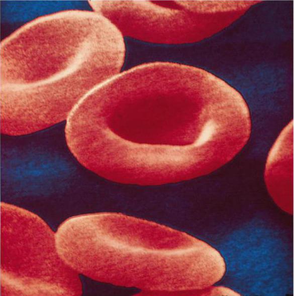
These blood cells are characterized by a biconcave shape and a red color due to the presence of a large amount of hemoglobin in the cell. It is hemoglobin that makes up the bulk of these cells. Their diameter varies from 7 to 8 microns, but the thickness reaches 2 - 2.5 microns. The nucleus in mature cells is absent, which significantly increases their surface. In addition, the absence of a core ensures rapid and uniform penetration of oxygen into the body. The life span of these cells is about 120 days. The total surface area of human red blood cells exceeds 3,000 square meters. This surface is 1500 times larger than the surface of the entire human body. If you place all the red cells of a person in one row, then you can get a chain, the length of which will be about 150,000 km. The destruction of these bodies occurs mainly in the spleen and partly in the liver.Functions
1. Nutritious: carry out the transfer of amino acids from the organs of the digestive system to the cells of the body;
2. Enzymatic:
are carriers of various enzymes ( specific protein catalysts);
3. Respiratory:
this function is carried out by hemoglobin, which is able to attach to itself and give off both oxygen and carbon dioxide;
4. Protective:
bind toxins due to the presence of special substances of protein origin on their surface.
Terms used to describe these cells
- microcytosis- the average size of red blood cells is less than normal;
- macrocytosis- the average size of red blood cells is larger than normal;
- normocytosis– the average size of red blood cells is normal;
- Anisocytosis- the sizes of red blood cells differ significantly, some are too small, others are very large;
- Poikilocytosis- the shape of the cells varies from regular to oval, sickle-shaped;
- Normochromia- red blood cells are colored normally, which is a sign of a normal level of hemoglobin in them;
- hypochromia- red blood cells are stained weakly, which indicates that they have less than normal hemoglobin.
Settling rate (ESR)
Erythrocyte sedimentation rate or ESR is a fairly well-known indicator of laboratory diagnostics, which means the rate of separation of unclotting blood, which is placed in a special capillary. Blood is divided into 2 layers - lower and upper. The bottom layer consists of settled red blood cells, but the top layer is plasma. This indicator is usually measured in millimeters per hour. The ESR value directly depends on the gender of the patient. In a normal state, in men, this indicator ranges from 1 to 10 mm / hour, but in women - from 2 to 15 mm / hour.With an increase in indicators, we are talking about violations of the body. There is an opinion that in most cases, ESR increases against the background of an increase in the ratio of large and small protein particles in the blood plasma. As soon as fungi, viruses or bacteria enter the body, the level of protective antibodies immediately increases, which leads to changes in the ratio of blood proteins. From this it follows that especially often ESR increases against the background of inflammatory processes such as inflammation of the joints, tonsillitis, pneumonia, etc. The higher this indicator, the more pronounced the inflammatory process. With a mild course of inflammation, the rate increases to 15 - 20 mm / h. If the inflammatory process is severe, then it jumps up to 60-80 mm/hour. If during the course of therapy the indicator begins to decrease, then the treatment was chosen correctly.
In addition to inflammatory diseases, an increase in ESR is also possible with some non-inflammatory ailments, namely:
- Malignant formations;
- Severe ailments of the liver and kidneys;
- Severe blood pathologies;
- Frequent blood transfusions;
- Vaccine therapy.
Hemolysis - what is it?
Hemolysis is the process of destruction of the membrane of red blood cells, as a result of which hemoglobin is released into the plasma and the blood becomes transparent.Modern experts distinguish the following types of hemolysis:
1. By the nature of the flow:
- Physiological: old and pathological forms of red cells are destroyed. The process of their destruction is noted in small vessels, macrophages ( cells of mesenchymal origin) bone marrow and spleen, as well as in liver cells;
- Pathological: against the background of a pathological condition, healthy young cells are destroyed.
- Endogenous: hemolysis occurs inside the human body;
- Exogenous: hemolysis occurs outside the body ( e.g. in a vial of blood).
- Mechanical: observed with mechanical ruptures of the membrane ( for example, a vial of blood had to be shaken);
- Chemical: observed when erythrocytes are exposed to substances that tend to dissolve lipids ( fatty substances) membranes. These substances include ether, alkalis, acids, alcohols and chloroform;
- Biological: noted when exposed to biological factors ( poisons of insects, snakes, bacteria) or transfusion of incompatible blood;
- Temperature: at low temperatures, ice crystals form in red blood cells, which tend to break the cell membrane;
- Osmotic: occurs when red blood cells enter an environment with a lower osmotic value than that of blood ( thermodynamic) pressure. Under this pressure, the cells swell and burst.
erythrocytes in the blood
 The total number of these cells in human blood is simply enormous. So, for example, if your weight is about 60 kg, then there are at least 25 trillion red blood cells in your blood. The figure is very large, so for practicality and convenience, experts do not calculate the total level of these cells, but their number in a small amount of blood, namely in its 1 cubic millimeter. It is important to note that the norms for the content of these cells are determined immediately by several factors - the age of the patient, his gender and place of residence.
 Norm of content of red blood cells
To determine the level of these cells helps clinical ( general) blood test .- In women - from 3.7 to 4.7 trillion in 1 liter;
- In men - from 4 to 5.1 trillion in 1 liter;
- In children over 13 years old - from 3.6 to 5.1 trillion per 1 liter;
- In children aged 1 to 12 years - from 3.5 to 4.7 trillion in 1 liter;
- In children at 1 year old - from 3.6 to 4.9 trillion in 1 liter;
- In children at six months - from 3.5 to 4.8 trillion per 1 liter;
- In children at 1 month - from 3.8 to 5.6 trillion in 1 liter;
- In children on the first day of their life - from 4.3 to 7.6 trillion in 1 liter.
The level of erythrocytes in the blood of pregnant women
Most often, the number of these bodies decreases slightly during pregnancy, which is completely normal. Firstly, during the gestation of the fetus, a large amount of water is retained in the woman's body, which enters the bloodstream and dilutes it. In addition, the organisms of almost all expectant mothers do not receive enough iron, as a result of which the formation of these cells again decreases.An increase in the level of red blood cells in the blood
A condition characterized by an increase in the level of red blood cells in the blood is called erythremia , erythrocytosis or polycythemia .The most common causes of this condition are:
- Polycystic kidney disease ( a disease in which cysts appear and gradually increase in both kidneys);
- COPD (chronic obstructive pulmonary disease - bronchial asthma, pulmonary emphysema, chronic bronchitis);
- Pickwick's syndrome ( obesity, accompanied by pulmonary insufficiency and arterial hypertension, i.e. persistent increase in blood pressure);
- Hydronephrosis ( persistent progressive expansion of the renal pelvis and calyx against the background of a violation of the outflow of urine);
- A course of steroid therapy;
- Congenital or acquired myeloma ( bone marrow tumors). A physiological decrease in the level of these cells is possible between 17.00 and 7.00, after eating and when taking blood in the supine position. You can find out about other reasons for lowering the level of these cells by consulting a specialist.
erythrocytes in urine
Normally, there should be no red blood cells in the urine. Their presence is allowed in the form of single cells in the field of view of the microscope. Being in the urine sediment in very small quantities, they may indicate that a person was involved in sports or did hard physical work. In women, a small amount of them can be observed with gynecological ailments, as well as during menstruation.A significant increase in their level in the urine can be noticed immediately, since the urine in such cases acquires a brown or red tint. The most common cause of the appearance of these cells in the urine is considered to be diseases of the kidneys and urinary tract. These include various infections, pyelonephritis ( inflammation of the kidney tissue), glomerulonephritis ( kidney disease characterized by inflammation of the glomerulus, ie. olfactory glomerulus), nephrolithiasis, and adenoma ( benign tumor) of the prostate gland. It is also possible to identify these cells in the urine with intestinal tumors, various blood clotting disorders, heart failure, smallpox ( contagious viral pathology), malaria ( acute infectious disease) etc.
Often, red blood cells appear in the urine and during therapy with certain medications such as urotropin. The fact of the presence of red blood cells in the urine should alert both the patient himself and his doctor. Such patients need a repeat urinalysis and a complete examination. A repeat urinalysis should be taken using a catheter. If the repeated analysis once again establishes the presence of numerous red cells in the urine, then the urinary system is already subjected to examination.
The erythrocyte, the structure and functions of which we will consider in our article, is the most important component of the blood. It is these cells that carry out gas exchange, providing respiration at the cellular and tissue level.
Erythrocyte: structure and functions
The circulatory system of humans and mammals is characterized by the most perfect structure compared to other organisms. It consists of a four-chambered heart and a closed system of blood vessels through which blood circulates continuously. This tissue consists of a liquid component - plasma, and a number of cells: erythrocytes, leukocytes and platelets. Every cell has a role to play. The structure of a human erythrocyte is determined by the functions performed. This concerns the size, shape and number of these blood cells.
Features of the structure of erythrocytes
Erythrocytes have the shape of a biconcave disc. They are not able to move independently in the bloodstream, like leukocytes. They reach the tissues and internal organs thanks to the work of the heart. Erythrocytes are prokaryotic cells. This means that they do not contain a decorated core. Otherwise, they could not carry oxygen and carbon dioxide. This function is performed due to the presence of a special substance inside the cells - hemoglobin, which also determines the red color of human blood.

The structure of hemoglobin
The structure and functions of erythrocytes are largely due to the characteristics of this particular substance. Hemoglobin has two components. This is an iron-containing component called heme, and a protein called globin. For the first time, the English biochemist Max Ferdinand Perutz managed to decipher the spatial structure of this chemical compound. For this discovery, he was awarded the Nobel Prize in 1962. Hemoglobin is a member of the group of chromoproteins. These include complex proteins consisting of a simple biopolymer and a prosthetic group. For hemoglobin, this group is heme. This group also includes plant chlorophyll, which ensures the flow of the process of photosynthesis.

How does gas exchange take place
In humans and other chordates, hemoglobin is located inside the red blood cells, while in invertebrates it is dissolved directly in the blood plasma. In any case, the chemical composition of this complex protein allows the formation of unstable compounds with oxygen and carbon dioxide. Oxygenated blood is called arterial blood. It is enriched with this gas in the lungs.
From the aorta, it goes to the arteries, and then to the capillaries. These smallest vessels are suitable for every cell of the body. Here, red blood cells give off oxygen and attach the main product of respiration - carbon dioxide. With the blood flow, which is already venous, they enter the lungs again. In these organs, gas exchange occurs in the smallest bubbles - alveoli. Here, hemoglobin removes carbon dioxide, which is removed from the body through exhalation, and the blood is again saturated with oxygen.
Such chemical reactions are due to the presence of ferrous iron in the heme. As a result of the connection and decomposition, oxy- and carbhemoglobin are sequentially formed. But the complex protein of erythrocytes can also form stable compounds. For example, incomplete combustion of fuel releases carbon monoxide, which forms carboxyhemoglobin with hemoglobin. This process leads to the death of red blood cells and poisoning of the body, which can lead to death.

What is anemia
Shortness of breath, noticeable weakness, tinnitus, noticeable pallor of the skin and mucous membranes may indicate an insufficient amount of hemoglobin in the blood. The norm of its content varies depending on the gender. In women, this figure is 120 - 140 g per 1000 ml of blood, and in men it reaches 180 g / l. The content of hemoglobin in the blood of newborns is the highest. It exceeds this figure in adults, reaching 210 g / l.
Lack of hemoglobin is a serious condition called anemia or anemia. It can be caused by a lack of vitamins and iron salts in foodstuffs, an addiction to alcohol, the effect of radiation pollution on the body and other negative environmental factors.
A decrease in the amount of hemoglobin may also be due to natural factors. For example, in women, anemia can be caused by the menstrual cycle or pregnancy. Subsequently, the amount of hemoglobin is normalized. A temporary decrease in this indicator is also observed in active donors who often donate blood. But an increased number of red blood cells is also quite dangerous and undesirable for the body. It leads to an increase in blood density and the formation of blood clots. Often an increase in this indicator is observed in people living in high mountainous areas.
It is possible to normalize the level of hemoglobin by eating foods containing iron. These include liver, tongue, meat of cattle, rabbit, fish, black and red caviar. Plant products also contain the necessary trace element, but the iron in them is much more difficult to digest. These include legumes, buckwheat, apples, molasses, red peppers and herbs.
Shape and size
The structure of blood erythrocytes is characterized primarily by their shape, which is quite unusual. It really resembles a disk concave on both sides. This form of red blood cells is not accidental. It increases the surface of red blood cells and ensures the most efficient penetration of oxygen into them. This unusual shape also contributes to an increase in the number of these cells. So, normally, 1 cubic mm of human blood contains about 5 million red blood cells, which also contributes to the best gas exchange.

The structure of frog erythrocytes
Scientists have long established that human red blood cells have structural features that provide the most efficient gas exchange. This applies to form, quantity, and internal content. This is especially evident when comparing the structure of human and frog erythrocytes. In the latter, red blood cells are oval in shape and contain a nucleus. This significantly reduces the content of respiratory pigments. Frog erythrocytes are much larger than human ones, and therefore their concentration is not so high. For comparison: if a person has more than 5 million of them in a cubic mm, then in amphibians this figure reaches 0.38.
Evolution of erythrocytes
The structure of human and frog erythrocytes allows us to draw conclusions about the evolutionary transformations of such structures. Respiratory pigments are also found in the simplest ciliates. In the blood of invertebrates, they are found directly in the plasma. But this significantly increases the density of the blood, which can lead to the formation of blood clots inside the vessels. Therefore, over time, evolutionary transformations went in the direction of the appearance of specialized cells, the formation of their biconcave shape, the disappearance of the nucleus, a decrease in their size and an increase in concentration.
Ontogenesis of red blood cells
The erythrocyte, the structure of which has a number of characteristic features, remains viable for 120 days. This is followed by their destruction in the liver and spleen. The main hematopoietic organ in humans is the red bone marrow. It continuously produces new red blood cells from stem cells. Initially, they contain a nucleus, which, as it matures, is destroyed and replaced by hemoglobin.
Features of blood transfusion
In a person's life, there are often situations in which a blood transfusion is required. For a long time, such operations led to the death of patients, and the real reasons for this remained a mystery. Only at the beginning of the 20th century it was established that the erythrocyte was to blame. The structure of these cells determines the blood groups of a person. There are four of them in total, and they are distinguished according to the AB0 system.
Each of them is distinguished by a special type of protein substances contained in red blood cells. They are called agglutinogens. They are absent in people with the first blood group. From the second - they have agglutinogens A, from the third - B, from the fourth - AB. At the same time, agglutinin proteins are contained in the blood plasma: alpha, beta, or both at the same time. The combination of these substances determines the compatibility of blood groups. This means that the simultaneous presence of agglutinogen A and agglutinin alpha in the blood is impossible. In this case, red blood cells stick together, which can lead to the death of the body.

What is the Rh factor
The structure of a human erythrocyte determines the performance of another function - the determination of the Rh factor. This sign is also necessarily taken into account during blood transfusion. In Rh-positive people, a special protein is located on the erythrocyte membrane. The majority of such people in the world - more than 80%. Rh-negative people do not have this protein.
What is the danger of mixing blood with red blood cells of different types? During the pregnancy of an Rh-negative woman, fetal proteins can enter her bloodstream. In response, the mother's body will begin to produce protective antibodies that neutralize them. During this process, the RBCs of the Rh-positive fetus are destroyed. Modern medicine has created special drugs that prevent this conflict.
Erythrocytes are red blood cells whose main function is to carry oxygen from the lungs to cells and tissues and carbon dioxide in the opposite direction. This role is possible due to the biconcave shape, small size, high concentration and the presence of hemoglobin in the cell.
Their main function is to transport oxygen (O2) from the lungs to the tissues and carbon dioxide (CO2) from the tissues to the lungs.
Mature erythrocytes do not have a nucleus and cytoplasmic organelles. Therefore, they are not capable of protein or lipid synthesis, ATP synthesis in the processes of oxidative phosphorylation. This sharply reduces the erythrocytes' own oxygen needs (no more than 2% of the total oxygen transported by the cell), and ATP synthesis is carried out during the glycolytic breakdown of glucose. About 98% of the mass of proteins in the erythrocyte cytoplasm is.
About 85% of red blood cells, called normocytes, have a diameter of 7-8 microns, a volume of 80-100 (femtoliters, or microns 3) and a shape - in the form of biconcave discs (discocytes). This provides them with a large gas exchange area (total for all erythrocytes is about 3800 m 2) and reduces the oxygen diffusion distance to the place of its binding to hemoglobin. Approximately 15% of erythrocytes have a different shape, size and may have processes on the surface of the cells.
Full-fledged "mature" erythrocytes have plasticity - the ability to reversibly deform. This allows them to pass through vessels with a smaller diameter, in particular, through capillaries with a lumen of 2-3 microns. This ability to deform is provided due to the liquid state of the membrane and the weak interaction between phospholipids, membrane proteins (glycophorins) and the cytoskeleton of intracellular matrix proteins (spectrin, ankyrin, hemoglobin). In the process of aging of erythrocytes, cholesterol and phospholipids with a higher content of fatty acids accumulate in the membrane, irreversible aggregation of spectrin and hemoglobin occurs, which causes a violation of the structure of the membrane, the shape of erythrocytes (they turn from discocytes into spherocytes) and their plasticity. Such red blood cells cannot pass through the capillaries. They are captured and destroyed by macrophages of the spleen, and some of them are hemolyzed inside the vessels. Glycophorins impart hydrophilic properties to the outer surface of erythrocytes and an electrical (zeta) potential. Therefore, erythrocytes repel each other and are in the plasma in a suspended state, determining the suspension stability of the blood.
Erythrocyte sedimentation rate (ESR)
Erythrocyte sedimentation rate (ESR)- an indicator characterizing the sedimentation of red blood cells when an anticoagulant (for example, sodium citrate) is added. The ESR is determined by measuring the height of the plasma column above the erythrocytes that have settled in a vertically located special capillary for 1 hour. The mechanism of this process is determined by the functional state of the erythrocyte, its charge, the protein composition of the plasma and other factors.
The specific gravity of erythrocytes is higher than that of blood plasma, therefore, in a capillary with blood, deprived of the ability to coagulate, they slowly settle. ESR in healthy adults is 1-10 mm/h in men and 2-15 mm/h in women. In newborns, the ESR is 1-2 mm/h, and in the elderly it is 1-20 mm/h.
The main factors affecting ESR include: the number, shape and size of red blood cells; the quantitative ratio of different types of blood plasma proteins; the content of bile pigments, etc. An increase in the content of albumins and bile pigments, as well as an increase in the number of erythrocytes in the blood, causes an increase in the zeta potential of cells and a decrease in ESR. An increase in the content of globulins, fibrinogen in the blood plasma, a decrease in the content of albumins and a decrease in the number of erythrocytes is accompanied by an increase in ESR.
One of the reasons for the higher ESR value in women compared to men is the lower number of red blood cells in the blood of women. ESR increases during dry eating and fasting, after vaccination (due to an increase in the content of globulins and fibrinogen in plasma), during pregnancy. A slowdown in ESR can be observed with an increase in blood viscosity due to increased evaporation of sweat (for example, under the action of high external temperature), with erythrocytosis (for example, in residents of high mountains or climbers, in newborns).
RBC count
The number of red blood cells in the peripheral blood of an adult is: in men - (3.9-5.1) * 10 12 cells / l; in women - (3.7-4.9). 10 12 cells/l. Their number in different age periods in children and adults is shown in Table. 1. In the elderly, the number of red blood cells approaches, on average, the lower limit of normal.
An increase in the number of erythrocytes per unit volume of blood above the upper limit of normal is called erythrocytosis: for men - above 5.1. 10 12 erythrocytes/l; for women - above 4.9. 10 12 erythrocytes/l. Erythrocytosis is relative and absolute. Relative erythrocytosis (without activation of erythropoiesis) is observed with an increase in blood viscosity in newborns (see Table 1), during physical work or exposure to high temperature. Absolute erythrocytosis is a consequence of enhanced erythropoiesis observed during human adaptation to high mountains or in endurance-trained individuals. Erygrocytosis develops with certain blood diseases (erythremia) or as a symptom of other diseases (heart or lung failure, etc.). With any type of erythrocytosis, the content of hemoglobin in the blood and hematocrit usually increase.
Table 1. Indicators of red blood in healthy children and adults
|
Erythrocytes 10 12 /l |
Reticulocytes, % |
Hemoglobin, g/l |
Hematocrit, % |
MCHC g/100 ml |
|||
|
newborns |
|||||||
|
1st week |
|||||||
|
6 months |
|||||||
|
grown men |
|||||||
|
adult women |
Note. MCV (mean corpuscular volume) - the average volume of erythrocytes; MCH (mean corpuscular hemoglobin) is the average content of hemoglobin in an erythrocyte; MCHC (mean corpuscular hemoglobin concentration) - hemoglobin content in 100 ml of erythrocytes (hemoglobin concentration in one erythrocyte).
erythropenia- This is a decrease in the number of red blood cells in the blood below the lower limit of normal. It can also be relative or absolute. Relative erythropenia is observed with an increase in fluid intake into the body with unchanged erythropoiesis. Absolute erythropenia (anemia) is a consequence of: 1) increased blood destruction (autoimmune hemolysis of erythrocytes, excessive blood-destroying function of the spleen); 2) a decrease in the effectiveness of erythropoiesis (with a deficiency of iron, vitamins (especially group B) in foods, the absence of an internal factor of Castle and insufficient absorption of vitamin B 12); 3) blood loss.
The main functions of red blood cells
transport function consists in the transfer of oxygen and carbon dioxide (respiratory or gas transport), nutrients (proteins, carbohydrates, etc.) and biologically active (NO) substances. Protective function erythrocytes lies in their ability to bind and neutralize certain toxins, as well as participate in blood clotting processes. Regulatory function erythrocytes lies in their active participation in maintaining the acid-base state of the body (blood pH) with the help of hemoglobin, which can bind CO 2 (thus reducing the content of H 2 CO 3 in the blood) and has ampholytic properties. Erythrocytes can also participate in the immunological reactions of the body, which is due to the presence in their cell membranes of specific compounds (glycoproteins and glycolipids) that have the properties of antigens (agglutinogens).
Life cycle of erythrocytes
The place of formation of red blood cells in the body of an adult is the red bone marrow. In the process of erythropoiesis, reticulocytes are formed from a pluripotent hematopoietic stem cell (PSHC) through a number of intermediate stages, which enter the peripheral blood and turn into mature erythrocytes after 24-36 hours. Their life span is 3-4 months. The place of death is the spleen (phagocytosis by macrophages up to 90%) or intravascular hemolysis (usually up to 10%).
Functions of hemoglobin and its compounds
The main functions of erythrocytes are due to the presence in their composition of a special protein -. Hemoglobin binds, transports and releases oxygen and carbon dioxide, providing the respiratory function of the blood, participates in the regulation, performing regulatory and buffer functions, and also gives red blood cells and blood a red color. Hemoglobin performs its functions only when it is in red blood cells. In the case of hemolysis of erythrocytes and the release of hemoglobin into the plasma, it cannot perform its functions. Plasma hemoglobin binds to the protein haptoglobin, the resulting complex is captured and destroyed by the cells of the phagocytic system of the liver and spleen. In massive hemolysis, hemoglobin is removed from the blood by the kidneys and appears in the urine (hemoglobinuria). Its elimination half-life is about 10 minutes.
The hemoglobin molecule has two pairs of polypeptide chains (globin is the protein part) and 4 hemes. Heme is a complex compound of protoporphyrin IX with iron (Fe 2+), which has a unique ability to attach or donate an oxygen molecule. At the same time, iron, to which oxygen is attached, remains divalent, it can easily be oxidized to trivalent as well. Heme is an active or so-called prosthetic group, and globin is a protein carrier of heme, creating a hydrophobic pocket for it and protecting Fe 2+ from oxidation.
There are a number of molecular forms of hemoglobin. The blood of an adult contains HbA (95-98% HbA 1 and 2-3% HbA 2) and HbF (0.1-2%). In newborns, HbF predominates (almost 80%), and in the fetus (up to 3 months of age) - hemoglobin type Gower I.
The normal content of hemoglobin in the blood of men averages 130-170 g/l, in women it is 120-150 g/l, in children it depends on age (see Table 1). The total hemoglobin content in the peripheral blood is approximately 750 g (150 g/L. 5 L of blood = 750 g). One gram of hemoglobin can bind 1.34 ml of oxygen. The optimal performance of the respiratory function by erythrocytes is noted with a normal content of hemoglobin in them. The content (saturation) of hemoglobin in an erythrocyte is reflected by the following indicators: 1) color index (CP); 2) MCH - the average content of hemoglobin in the erythrocyte; 3) MCHC - the concentration of hemoglobin in the erythrocyte. Erythrocytes with normal hemoglobin content are characterized by CP = 0.8-1.05; MCH = 25.4-34.6 pg; MCHC = 30-37 g/dl and are called normochromic. Cells with reduced hemoglobin content have CP< 0,8; МСН < 25,4 пг; МСНС < 30 г/дл и получили название гипохромных. Эритроциты с повышенным содержанием гемоглобина (ЦП >1.05; MSI > 34.6 pg; MCHC > 37 g/dl) are called hyperchromic.
The cause of erythrocyte hypochromia is most often their formation in conditions of iron deficiency (Fe 2+) in the body, and hyperchromia - in conditions of a lack of vitamin B 12 (cyanocobalamin) and (or) folic acid. In a number of regions of our country, there is a low content of Fe 2+ in water. Therefore, their inhabitants (especially women) are more likely to develop hypochromic anemia. For its prevention, it is necessary to compensate for the lack of iron intake with water with food products containing it in sufficient quantities or with special preparations.
Hemoglobin compounds
Hemoglobin bound to oxygen is called oxyhemoglobin (HbO2). Its content in arterial blood reaches 96-98%; HbO 2, which gave up O 2 after dissociation, is called reduced (HHb). Hemoglobin binds carbon dioxide, forming carbhemoglobin (HbCO 2). The formation of HbCO 2 not only promotes the transport of CO 2 , but also reduces the formation of carbonic acid and thus maintains the bicarbonate buffer of the blood plasma. Oxyhemoglobin, reduced hemoglobin and carbhemoglobin are called physiological (functional) compounds of hemoglobin.
Carboxyhemoglobin is a compound of hemoglobin with carbon monoxide (CO - carbon monoxide). Hemoglobin has a significantly greater affinity for CO than for oxygen, and forms carboxyhemoglobin at low concentrations of CO, while losing the ability to bind oxygen and endangering life. Another non-physiological compound of hemoglobin is methemoglobin. In it, iron is oxidized to a trivalent state. Methemoglobin is not able to enter into a reversible reaction with O 2 and is a functionally inactive compound. With its excessive accumulation in the blood, a threat to human life also arises. In this regard, methemoglobin and carboxyhemoglobin are also called pathological hemoglobin compounds.
In a healthy person, methemoglobin is constantly present in the blood, but in very small quantities. The formation of methemoglobin occurs under the action of oxidizing agents (peroxides, nitro derivatives of organic substances, etc.), which constantly enter the blood from the cells of various organs, especially the intestines. The formation of methemoglobin is limited by antioxidants (glutathione and ascorbic acid) present in erythrocytes, and its reduction to hemoglobin occurs during enzymatic reactions involving erythrocyte dehydrogenase enzymes.
Erythropoiesis
Erythropoiesis - is the process of formation of red blood cells from PSGC. The number of erythrocytes contained in the blood depends on the ratio of erythrocytes formed and destroyed in the body at the same time. In a healthy person, the number of formed and destroyed erythrocytes is equal, which ensures the maintenance of a relatively constant number of erythrocytes in the blood under normal conditions. The totality of body structures, including peripheral blood, organs of erythropoiesis and destruction of erythrocytes, is called erythrone.
In a healthy adult, erythropoiesis occurs in the hematopoietic space between the sinusoids of the red bone marrow and ends in the blood vessels. Under the influence of signals from microenvironment cells activated by the destruction products of erythrocytes and other blood cells, early-acting PSGC factors differentiate into committed oligopotent (myeloid) and then into unipotent hematopoietic stem cells of the erythroid series (BFU-E). Further differentiation of erythroid cells and the formation of the immediate precursors of erythrocytes - reticulocytes occurs under the influence of late-acting factors, among which the hormone erythropoietin (EPO) plays a key role.
Reticulocytes enter the circulating (peripheral) blood and are converted into red blood cells within 1-2 days. The content of reticulocytes in the blood is 0.8-1.5% of the number of red blood cells. The lifespan of red blood cells is 3-4 months (average 100 days), after which they are removed from the bloodstream. About (20-25) is replaced in the blood per day. 10 10 erythrocytes by reticulocytes. The efficiency of erythropoiesis in this case is 92-97%; 3-8% of erythrocyte precursor cells do not complete the differentiation cycle and are destroyed in the bone marrow by macrophages - ineffective erythropoiesis. Under special conditions (for example, stimulation of erythropoiesis in anemia), ineffective erythropoiesis can reach 50%.
Erythropoiesis depends on many exogenous and endogenous factors and is regulated by complex mechanisms. It depends on sufficient intake of vitamins, iron, other trace elements, essential amino acids, fatty acids, protein and energy in the body with food. Their insufficient intake leads to the development of alimentary and other forms of deficiency anemia. Among the endogenous factors regulating erythropoiesis, the leading place is given to cytokines, especially erythropoietin. EPO is a glycoprotein hormone and the main regulator of erythropoiesis. EPO stimulates the proliferation and differentiation of all erythrocyte precursor cells, starting with BFU-E, increases the rate of hemoglobin synthesis in them and inhibits their apoptosis. In an adult, the main site of EPO synthesis (90%) is the peritubular cells of the night, in which the formation and secretion of the hormone increase with a decrease in oxygen tension in the blood and in these cells. Synthesis of EPO in the kidneys is enhanced under the influence of growth hormone, glucocorticoids, testosterone, insulin, norepinephrine (through stimulation of β1-adrenergic receptors). EPO is synthesized in small amounts in liver cells (up to 9%) and bone marrow macrophages (1%).
In the clinic, recombinant erythropoietin (rHuEPO) is used to stimulate erythropoiesis.
The female sex hormones estrogens inhibit erythropoiesis. The nervous regulation of erythropoiesis is carried out by the ANS. At the same time, an increase in the tone of the sympathetic section is accompanied by an increase in erythropoiesis, and the parasympathetic section is accompanied by a weakening.
Table of contents of the subject "Functions of Blood Cells. Erythrocytes. Neutrophils. Basophils.":1. Functions of blood cells. Functions of erythrocytes. properties of erythrocytes. Embden-Meyerhof cycle. The structure of erythrocytes.
2. Hemoglobin. Types (types) of hemoglobin. Synthesis of hemoglobin. hemoglobin function. The structure of hemoglobin.
3. Aging of erythrocytes. Destruction of erythrocytes. The lifespan of an erythrocyte. Echinocyte. Echinocytes.
4. Iron. Iron is normal. The role of iron ions in erythropoiesis. Transferrin. The body's need for iron. iron deficiency. OZHSS.
5. Erythropoiesis. erythroblastic islets. Anemia. Erythrocytosis.
6. Regulation of erythropoiesis. Erythropoietin. Sex hormones and erythropoiesis.
7. Leukocytes. Leukocytosis. Leukopenia. Granulocytes. Leukocyte formula.
8. Functions of neutrophilic granulocytes (leukocytes). Defensins. Cathelicidins. Acute phase proteins. chemotactic factors.
9. Bactericidal effect of neutrophils. Granulopoiesis. Neutrophilic granulopoiesis. Granulocytosis. Neutropenia.
10. Functions of basophils. Functions of basophilic granulocytes. Normal amount. Histamine. Heparin.
Functions of blood cells. Functions of erythrocytes. properties of erythrocytes. Embden-Meyerhof cycle. The structure of erythrocytes.
Whole blood consists of a liquid part (plasma) and formed elements, which include erythrocytes, leukocytes and platelets - platelets.
Blood functions:
1) transport- transfer of gases (02 and CO2), plastic (amino acids, nucleosides, vitamins, minerals), energy (glucose, fats) resources to tissues, and end products of metabolism - to excretory organs (gastrointestinal tract, lungs, kidneys, sweat glands, skin);
2) homeostatic- maintenance of body temperature, acid-base state of the body, water-salt metabolism, tissue homeostasis and tissue regeneration;
3) protective- providing immune responses, blood and tissue barriers against infection;
4) regulatory- humoral and hormonal regulation of the functions of various systems and tissues;
5) secretory- the formation of biologically active substances by blood cells.
Functions and properties of erythrocytes
red blood cells carry 02 contained in hemoglobin from the lungs to tissues and CO2 from tissues to the alveoli of the lungs. The functions of erythrocytes are due to the high content of hemoglobin (95% of the erythrocyte mass), the deformability of the cytoskeleton, due to which erythrocytes easily penetrate through capillaries with a diameter of less than 3 microns, although they have a diameter of 7 to 8 microns. Glucose is the main source of energy in the erythrocyte. Restoration of the shape of an erythrocyte deformed in the capillary, active membrane transport of cations through the erythrocyte membrane, and synthesis of glutathione are provided by the energy of anaerobic glycolysis in Embden-Meyerhof cycle. During the metabolism of glucose in erythrocyte along the side pathway of glycolysis, controlled by the enzyme diphosphoglycerate mutase, 2,3-diphosphoglycerate (2,3-DPG) is formed in the erythrocyte. The main value of 2,3-DFG is to reduce the affinity of hemoglobin for oxygen.
AT Embden-Meyerhof cycle 90% of the glucose consumed by red blood cells is consumed. Inhibition of glycolysis, which occurs, for example, during aging of the erythrocyte and reduces the concentration of ATP in the erythrocyte, leads to the accumulation of sodium and water ions, calcium ions in it, damage to the membrane, which reduces mechanical and osmotic stability erythrocyte, and aging erythrocyte is destroyed. The energy of glucose in the erythrocyte is also used in reduction reactions that protect the components erythrocyte from oxidative denaturation that impairs their function. Due to the reduction reactions, the iron atoms of hemoglobin are maintained in a reduced, i.e., divalent form, which prevents the conversion of hemoglobin into methemoglobin, in which iron is oxidized to trivalent, as a result of which methemoglobin is unable to transport oxygen. Restoration of oxidized iron methemoglobin to divalent is provided by the enzyme - methemoglobin reductase. In the reduced state, sulfur-containing groups included in the erythrocyte membrane, hemoglobin, and enzymes are also maintained, which preserves the functional properties of these structures.
red blood cells have a disc-shaped, biconcave shape, their surface is about 145 µm2, and the volume reaches 85-90 µm3. Such a ratio of area to volume contributes to the deformability (the latter is understood as the ability of erythrocytes to reversible changes in size and shape) of erythrocytes during their passage through the capillaries. The shape and deformability of erythrocytes are maintained by membrane lipids - phospholipids (glycerophospholipids, sphingolipids, phosphatidylethanolamine, phosphatidylsyrin, etc.), glycolipids and cholesterol, as well as proteins of their cytoskeleton. The composition of the cytoskeleton erythrocyte membrane proteins are included spectrin(major cytoskeletal protein), ankyrin, actin, band proteins 4.1, 4.2, 4.9, tropomyosin, tropomodulin, adzucin. The basis of the erythrocyte membrane is a lipid bilayer penetrated by integral proteins of the cytoskeleton - glycoproteins and band 3 protein. The latter are associated with a part of the cytoskeletal protein network - the spectrin-actin-band 4.1 protein complex, localized on the cytoplasmic surface of the lipid bilayer erythrocyte membrane(Fig. 7.1).
The interaction of the protein cytoskeleton with the lipid bilayer of the membrane ensures the stability of the structure of the erythrocyte, the behavior of the erythrocyte as an elastic solid body during its deformation. Non-covalent intermolecular interactions of cytoskeletal proteins easily provide a change in the size and shape of erythrocytes (their deformation) when these cells pass through the microvasculature, when reticulocytes exit the bone marrow into the blood - due to a change in the location of spectrin molecules on the inner surface of the lipid bilayer. Genetic abnormalities of cytoskeletal proteins in humans are accompanied by defects in the erythrocyte membrane. As a result, the latter acquire an altered form (the so-called spherocytes, elliptocytes, etc.) and have an increased tendency to hemolysis. An increase in the ratio of cholesterol-phospholipids in the membrane increases its viscosity, reduces the fluidity and elasticity of the erythrocyte membrane. As a result, the deformability of the erythrocyte decreases. Increased oxidation of unsaturated fatty acids of membrane phospholipids by hydrogen peroxide or superoxide radicals causes hemolysis of erythrocytes ( destruction of red blood cells with the release of hemoglobin into the environment), damage to the erythrocyte hemoglobin molecule. Glutathione constantly formed in the erythrocyte, as well as antioxidants (ostocopherol), enzymes - glutathione reductase, superoxide dismutase, etc. protect the components of the erythrocyte from this damage.
 Rice. 7.1. Scheme of the model of changes in the cytoskeleton of the erythrocyte membrane during its reversible deformation. Reversible deformation of the erythrocyte changes only the spatial configuration (stereometry) of the erythrocyte, following the change in the spatial arrangement of the cytoskeleton molecules. With these changes in the shape of the erythrocyte, the surface area of the erythrocyte remains unchanged. a - the position of the molecules of the cytoskeleton of the erythrocyte membrane in the absence of its deformation. Spectrin molecules are in a collapsed state.
Up to 52% mass erythrocyte membranes proteins are glycoproteins, which form blood group antigens with oligosaccharides. Membrane glycoproteins contain sialic acid, which gives a negative charge to red blood cells, repelling them from each other.
membrane enzymes- Ka+/K+-dependent ATPase provides active transport of Na+ from the erythrocyte and K+ into its cytoplasm. Ca2+-dependent ATPase removes Ca2+ from the erythrocyte. The erythrocyte enzyme carbonic anhydrase catalyzes the reaction: Ca2 + H20 H2CO3 o H + + HCO3, therefore, the erythrocyte transports part of the carbon dioxide from the tissues to the lungs in the form of bicarbonate, up to 30% CO2 is transported by the hemoglobin of erythrocytes in the form of a carbamic compound with the globin NH2 radical.
It consists in the transport of various substances by the blood. A specific feature of blood is the transport of O 2 and CO 2. The transport of gases is carried out by erythrocytes and plasma.
characteristics of erythrocytes.(Er).
The form: 85% Er is a biconcave disc, easily deformed, which is necessary for its passage through the capillary. Erythrocyte diameter = 7.2 - 7.5 µm.
More than 8 microns - macrocytes.
Less than 6 microns - microcytes.
Quantity:
M - 4.5 - 5.0 ∙ 10 12 / l. . - erythrocytosis.
F - 4.0 - 4.5 ∙ 10 12 / l. ↓ - erythropenia.
Membrane Er easily permeable for anions HCO 3 - Cl, as well as for O 2, CO 2, H +, OH -.
Hardly permeable for K + , Na + (1 million times lower than for anions).
properties of erythrocytes.
1) Plasticity- the ability to reversible deformation. As we age, this ability decreases.
The transformation of Er into spherocytes leads to the fact that they cannot pass through the capillary and are retained in the spleen and phagocytosed.
Plasticity depends on the properties of the membrane and the properties of hemoglobin, on the ratio of different lipid fractions in the membrane. Especially important is the ratio of phospholipids and cholesterol, which determine the fluidity of membranes.
This ratio is expressed as a lipolytic coefficient (LC):
Normal LA = cholesterol / lecithin = 0.9
↓ cholesterol → ↓ membrane stability, fluidity property changes.
Lecithin → erythrocyte membrane permeability.
2) Osmotic stability of the erythrocyte.
R osm. in the erythrocyte is higher than in plasma, which provides cell turgor. It is created by a high intracellular concentration of proteins, more than in plasma. In a hypotonic solution, the Er swell, in a hypertonic solution they shrink.
3) Ensuring creative connections.
Various substances are transported on the erythrocyte. This provides intercellular communication.
It has been shown that when the liver is damaged, erythrocytes begin to intensively transport nucleotides, peptides, and amino acids from the bone marrow to the liver, contributing to the restoration of the structure of the organ.
4) The ability of erythrocytes to settle.
Albumins- lyophilic colloids, create a hydrated shell around the erythrocyte and keep them in suspension.
Globulins – lyophobic colloids- reduce the hydration shell and the negative surface charge of the membrane, which contributes to increased erythrocyte aggregation.
The ratio of albumins and globulins is the protein coefficient of BC. Fine
BC \u003d albumins / globulins \u003d 1.5 - 1.7
With a normal protein coefficient of ESR in men, 2 - 10 mm / hour; in women 2 - 15 mm / hour.
5) Aggregation of erythrocytes.
With a slowdown in blood flow and an increase in blood viscosity, erythrocytes form aggregates that lead to rheological disorders. This happens:
1) with traumatic shock;
2) postinfarction collapse;
3) peritonitis;
4) acute intestinal obstruction;
5) burns;
5) acute pancreatitis and other conditions.
6) Destruction of erythrocytes.
The lifespan of an erythrocyte in the bloodstream is ~ 120 days. During this period, the physiological aging of the cell develops. About 10% of erythrocytes are destroyed normally in the vascular bed, the rest in the liver, spleen.
Functions of erythrocytes.
1) Transport of O 2 , CO 2 , AA, peptides, nucleotides to various organs for regenerative processes.
2) The ability to adsorb toxic products of endogenous and exogenous, bacterial and non-bacterial origin and inactivate them.
3) Participation in the regulation of blood pH due to the hemoglobin buffer.
4) Er. take part in blood coagulation and fibrinolysis, sorbing factors of the coagulation and anticoagulation systems on the entire surface.
5) Er. participate in immunological reactions, such as agglutination, because their membranes contain antigens - agglutinogens.
Functions of hemoglobin.
Found in erythrocytes. The share of hemoglobin accounts for 34% of the total and 90 - 95% of the dry mass of the erythrocyte. It provides O 2 and CO 2 transport. This is a chromoprotein. It consists of 4 iron-containing heme groups and a globin protein residue. Iron Fe 2+.
M. from 130 to 160 g / l (cf. 145 g / l).
F. from 120 to 140g/l.
Hb synthesis begins in normocytes. As the erythroid cell matures, Hb synthesis decreases. Mature erythrocytes do not synthesize Hb.
The process of Hb synthesis during erythropoiesis is associated with the consumption of endogenous iron.
With the destruction of red blood cells from hemoglobin, the bile pigment bilirubin is formed, which in the intestine turns into stercobilin, and in the kidneys into urobilin and is excreted with feces and urine.
Types of hemoglobin.
7 - 12 weeks of intrauterine development - Hv R (primitive). At the 9th week - Hb F (fetal). By the time of birth, Nv A appears.
During the first year of life, Hb F is completely replaced by Hb A.
Hb P and Hb F have a higher affinity for O 2 than Hb A, i.e., the ability to saturate with O 2 with a lower content in the blood.
Affinity is determined by globins.
Compounds of hemoglobin with gases.
The combination of hemoglobin with oxygen, called oxyhemoglobin (HbO 2 ), provides the scarlet color of arterial blood.
Blood oxygen capacity (KEK).
This is the amount of oxygen that 100g of blood can bind. It is known that one g of hemoglobin binds 1.34 ml of O 2 . KEK \u003d Hb ∙ 1.34. For arterial blood cake = 18 - 20 vol% or 180 - 200 ml/l of blood.
Oxygen capacity depends on:
1) the amount of hemoglobin.
2) blood temperature (when heated, the blood decreases)
3) pH (decreases when acidified)
Pathological compounds of hemoglobin with oxygen.
Under the action of strong oxidizing agents, Fe 2+ turns into Fe 3+ - this is a strong compound of methemoglobin. When it accumulates in the blood, death occurs.
Compounds of hemoglobin with CO 2
called carbhemoglobin HBCO 2 . In arterial blood it contains 52% or 520 ml/l. In the venous - 58% or 580 ml / l.
The pathological combination of hemoglobin with CO is called carboxyhemoglobin (HbCO). The presence of even 0.1% CO2 in the air converts 80% of hemoglobin into carboxyhemoglobin. The connection is stable. Under normal conditions, it decomposes very slowly.
Help with carbon monoxide poisoning.
1) provide oxygen access
2) inhalation of pure oxygen increases the rate of decomposition of carboxyhemoglobin by 20 times.
Myoglobin.
This is hemoglobin contained in the muscles and myocardium. Provides oxygen demand during contraction with cessation of blood flow (static tension of skeletal muscles).
Erythrokinetics.
This is understood as the development of erythrocytes, their functioning in the vascular bed and destruction.
Erythropoiesis
Hemocytopoiesis and erythropoiesis occur in myeloid tissue. The development of all shaped elements comes from a pluripotent stem cell.
KPL → SK → CFU ─GEMM
KPT- l KPV- l N E B
Factors affecting stem cell differentiation.
1. Lymphokines. They are secreted by leukocytes. Many lymphokines - a decrease in differentiation towards the erythroid series. A decrease in the content of lymphokines - an increase in the formation of red blood cells.
2. The main stimulant of erythropoiesis is the oxygen content in the blood. A decrease in the content of O 2 , a chronic deficiency of O 2 are a system-forming factor that is perceived by central and peripheral chemoreceptors. The chemoreceptor of the juxtaglomerular complex of the kidney (JGCC) is important. It stimulates the formation of erythropoietin, which increases:
1) stem cell differentiation.
2) accelerates the maturation of erythrocytes.
3) accelerates the release of erythrocytes from the bone marrow depot

In this case, there is true(absolute)erythrocytosis. The number of red blood cells in the body increases.
False erythrocytosis occurs when there is a temporary decrease in oxygen in the blood
(for example, during physical work). In this case, erythrocytes leave the depot and their number grows only in a unit of blood volume, but not in the body.
Erythropoiesis
The formation of erythrocytes occurs when erythroid cells interact with bone marrow macrophages. These cell associations are called erythroblastic islands (EOs).
EO macrophages influence erythrocyte proliferation and maturation by:
1) phagocytosis of nuclei ejected by the cell;
2) receipt of ferritin and other plastic materials from the macrophage into erythroblasts;
3) secretion of erythropoietin active substances;
4) creating favorable conditions for the development of erythroblasts.
RBC formation
200 - 250 billion erythrocytes are formed per day
proerythroblast (doubling).
2
basophilic
4 basophilic EB II order.
8 polychromatic erythroblasts of the first order.
polychromatophilic
↓16 polychromatophilic erythroblasts of the second order.
32 PCP normoblasts.
3
oxyphilic
32 reticulocytes.
32 erythrocytes.
Factors necessary for the formation of an erythrocyte.
1) Iron needed for gem synthesis. 95% of the daily requirement is received by the body from collapsing red blood cells. 20 - 25 mg of Fe is required daily.
iron depot.
1) Ferritin- in macrophages in the liver, intestinal mucosa.
2) Hemosiderin- in the bone marrow, liver, spleen.
Iron stores are needed for an emergency change in the synthesis of red blood cells. Fe in the body is 4 - 5g, of which ¼ is reserve Fe, the rest is functional. 62 - 70% of it is in the composition of red blood cells, 5 - 10% in myoglobin, the rest in tissues, where it participates in many metabolic processes.
In the bone marrow, Fe is taken up predominantly by basophilic and polychromatophilic pronormoblasts.
Iron is delivered to erythroblasts in combination with a plasma protein, transferrin.
In the gastrointestinal tract, iron is better absorbed in the 2-valence state. This state is supported by ascorbic acid, fructose, AA - cysteine, methionine.
Iron, which is part of gemma (in meat products, blood sausages), is better absorbed in the intestine than iron from plant products. 1 μg is absorbed daily.
The role of vitamins
AT 12 - an external hematopoietic factor (for the synthesis of nucleoproteins, maturation and division of cell nuclei).
With a deficiency of B 12, megaloblasts are formed, of which megalocytes with a short lifespan. The result is anemia. Reason B 12 - deficiency - lack of intrinsic factor Castle (glycoprotein that binds B 12 , protects B 12 from digestion by digestive enzymes). Castle factor deficiency is associated with atrophy of the gastric mucosa, especially in the elderly. Stocks B 12 for 1 to 5 years, but its depletion leads to disease.
B 12 is found in the liver, kidneys, eggs. The daily requirement is 5 mcg.
Folic acid DNA, globin (supports DNA synthesis in bone marrow cells and globin synthesis).
The daily requirement is 500 - 700 mcg, there is a reserve of 5 - 10 mg, a third of it is in the liver.
Lack of B 9 - anemia associated with accelerated destruction of red blood cells.
Found in vegetables (spinach), yeast, milk.
AT 6 - pyridoxine - for the formation of heme.
AT 2 - for the formation of stroma, deficiency causes anemia of the hyporegenerative type.
Pantothenic acid - the synthesis of phospholipids.
Vitamin C - supports the main stages of erythropoiesis: the metabolism of folic acid, iron, (heme synthesis).
Vitamin E - protects the phospholipids of the erythrocyte membrane from peroxidation, which enhances the hemolysis of erythrocytes.
RR - too.
trace elements Ni, Co, selenium cooperates with vitamin E, Zn - 75% of it is in erythrocytes as part of carbonic anhydrase.
Anemia:
1) due to a decrease in the number of red blood cells;
2) decrease in hemoglobin content;
3) both reasons together.
Stimulation of erythropoiesis occurs under the influence of ACTH, glucocorticoids, TSH,
catecholamines through β - AR, androgens, prostaglandins (PGE, PGE 2), sympathetic system.
brakes an inhibitor of erythropoiesis during pregnancy.
Anemia
1) due to a decrease in the number of red blood cells
2) a decrease in the amount of hemoglobin
3) both reasons together.
Functioning of erythrocytes in the vascular bed
The quality of the functioning of red blood cells depends on:
1) erythrocyte size
2) forms of erythrocyte
3) the type of hemoglobin in erythrocytes
4) the amount of hemoglobin in erythrocytes
4) the number of erythrocytes in peripheral blood. This is related to the work of the depot.
RBC destruction
They live a maximum of 120 days, on average 60 - 90.
With aging, the production of ATP decreases during glucose metabolism. This results in:
1) to a violation of the ionic composition of the contents of the erythrocyte. As a result - osmotic hemolysis in the vessel;
2) The lack of ATP leads to a violation of the elasticity of the erythrocyte membrane and causes mechanical hemolysis in the vessel;
In intravascular hemolysis, hemoglobin is released into the plasma, binds to plasma haptoglobin, and leaves the plasma to be absorbed by the liver parenchyma.