
Acquired cataract in children. Causes, symptoms, methods of treatment of congenital and other types of cataracts in children
One of the most common eye diseases is cataract. It is mainly diagnosed in adults and the elderly, but it can also be found in children.
The prevalence in newborns is 5 people per 100 thousand, in older children - 3-4 cases per 10 thousand people.
Disease Definition
Cataract is an eye disease in which there is a clouding of the lens substance with partial or complete loss of sharpness and clarity of vision. Turbidity can be both total and incomplete.
According to the International Classification of Diseases 10th revision, nosology is coded as H25-H28. But a congenital disease in children according to ICD-10 has the code Q12.0.
The lens is a biconvex lens, it refracts the sun's rays passing through it and focuses them on the retina.
Irritation from the retina is transmitted along the optic nerve to the information processing areas in the brain.
With cataracts, due to turbidity, the refraction of sunlight is disturbed, the image becomes blurry.
Etiology
It is not possible to find the exact cause of cataracts, but there are factors that may predispose to its development:
The leading factor in the appearance of a congenital form of cataract is heredity. Often, among close relatives of a sick child (mother, father, brothers and sisters), there are cases of cataracts in history.
The disease is linked to some genes, there is a high probability of cataracts in offspring.
Causes of congenital pathology in children:
But congenital cataract is registered and in children without burdened heredity. How can this be explained?
The fetus is very susceptible to viral infections during the first trimester of pregnancy.
If at this time he is attacked by viruses, then the congenital form may develop and become the least of the evils that viruses can inflict on the fetus.
Causative agents of intrauterine infection:

In diabetes, there is an increase in the glucose content in the lens due to hyperglycemia. The lens fibers swell, lose their transparency - this is how this type of cataract begins.
With galactosemia, the accumulation of galactose in the lens similarly occurs. In transmitted light, it looks like oil droplets. These accumulations can be seen already during the first days of a child's life.
In traumatic lesions, regardless of age rosette cataracts develop, which progress and can completely occupy the entire lens.
Opacification of the lens can occur as a complication of other diseases. For example, in uveitis, inflammatory products can enter the lens, leading to the development of cataracts.
Various radiations have a negative effect on the lens: infrared, ultraviolet. There is a peeling of the anterior chamber of the lens, which leads to its clouding.
With a deficiency of calcium ions in the body, calcium cataracts occur. Its development is possible with the removal of the parathyroid glands responsible for calcium metabolism.
Turbidity appears as small, sometimes bright dots on the pupil, which can be seen with the naked eye. Treatment of children with punctate cataract is long.
The constant use of certain drugs can also lead to illness. The list includes hormonal drugs, cardiac glycosides.
The ingestion of various substances, such as alkalis, leads to toxic cataracts. Alkali reduces the acidity of the anterior chamber of the eye, glucose is washed out of the lens.
Causes, symptoms and treatment of the disease:
Classification
Depending on the age of occurrence of cataracts, 2 types of cataracts are distinguished - congenital and acquired.
More often, ophthalmologists encounter acquired cataracts, congenital cataracts are quite rare.
Depending on the stage, there are:
- initial;
- immature;
- mature;
- overripe.

Clinical manifestations
A newborn baby has a cataract usually at periodic medical examinations - you should not avoid them. You can suspect a cataract in a child on your own in the following cases:
- the child practically does not react to silent toys;
- does not accompany the gaze of parents - does not focus vision;
- rapid uncontrolled eye movements;
- gray or white pupil.
- examination and assessment of the condition;
- operation;
- rehabilitation.
- chromosomal abnormalities: trisomy 21 chromosomes, Turner syndrome, trisomy 13 chromosomes, trisomy 18 pairs, cat's cry syndrome;
- craniofacial syndromes: cerebro-oculofacial skeletal syndrome, nephropathy, Lowe's syndrome, Allport's syndrome, Hallermann-Streiff-Francois syndrome;
- skeletopathies: Smith-Lemli-Opitz syndrome, Conradi syndrome, Weill-Marquezani syndrome, Stickler syndrome, Bardet-Biedl syndrome, Rubinstein-Taybi syndrome, syndactyly and polydactyly;
- neurometabolic diseases: Zellweger syndrome, Meckel-Gruber syndrome, Marinescu-Sjogren syndrome, infantile neuronal lipofuscinosis;
- myopathies: myotonic dystrophy;
- dermatological: crystalline cataract and non-combable hair, Cockayne syndrome, Rothmund-Thomson syndrome, atopic dermatitis, pigment incontinence syndrome, progeria, congenital ichthyosis, ectodermal dysplasia, Werner's syndrome.
- intrauterine infection (, toxoplasmosis);
- infection acquired at an early age, for example Herpes Simlex(cataract as a result of uveitis or parsplanitis);
- medicinal (corticosteroids, chlorpromazine);
- metabolic disorders (galactosemia, galactokinase deficiency, hypocalcemia, hypoglycemia, diabetes mellitus, mannosidosis, hyperferritinemia);
- trauma (accident or intentionally inflicted injury, laser coagulation, radiation injury);
- due to other diseases: endophthalmitis, retinopathy of prematurity, persistent primary vitreous, retinitis pigmentosa, aniridia, microphthalmia).
- manifesting immediately after birth (congenital) or acquired over the course of life;
- hereditary and non-hereditary.
- fetal nuclear - clouding of the lens substance between the anterior and posterior Y-shaped sutures, which is often accompanied by clouding of the posterior capsule;
- cortical - clouding of the anterior or posterior cortical layers, not extending to the fetal nucleus and often accompanied by clouding of the posterior capsule;
- persistent fetal vasculature - the presence of one or more of the listed signs (retrolental membrane with or without visible vessels, passable or impassable hyaloid vessel, stretched ciliary processes);
- isolated opacification of the posterior capsule (posterior polar cataract) - opacification of the posterior capsule without opacification of the layers of the cortex that lie closer to the center and the nucleus;
- posterior lentiglobus - protrusion of the posterior capsule posteriorly with or without a defect in the posterior capsule.
- partial;
- complete (total - the entire lens is diffusely cloudy).
- Unilateral - rarely accompanied by a systemic disease. Persistent fetal vasculature is a common cause of unilateral cataract. Another common cause of this type of cataract in children is trauma. May develop after laser coagulation for retinopathy of prematurity or after intraocular surgery.
- Bilateral - the etiology is established in approximately 50% of children. The most common cause of this form in Europe and the United States is an autosomal dominant hereditary cataract.
- With myopic refraction and astigmatism, the most complete correction is prescribed.
- With hypermetropia, the prescribed correction depends on the age of the child. Up to a year, weak myopic refraction is formed with glasses.
- From two years of age and older, two pairs of distance and near glasses, or one bifocal, or complex progressive glasses are assigned.
- direct - the better seeing eye is closed (it is prescribed for a period of several hours a day to a month, depending on the severity of amblyopia);
- alternately direct - the leading eye closes more often (the ratio of closing in time is 1:2, 1:3, 1:6, and so on).
- glare (general glare according to Bangerter) - a large area of the retina is illuminated, its inhibition develops, and the area of the macula is the first to come out of inhibition, since bright light is an adequate irritant for cones. After the illumination, the child is asked to perform a close visual load for 5-10 minutes;
- maculotester device for the formation of the correct fixation;
- sensory training (computer pleoptics);
- physiotherapy (laser stimulation, magnetic stimulation).
- in the first year, the inspection is carried out once every two months;
- for the second and third years - once every 3-4 months;
- in the future - once every six months.
- metabolic disorders in the body;
- infections during fetal development;
- chromosomal diseases (Down syndrome and others);
- congenital predisposition to the pathology of the lens;
- the use by a pregnant woman of drugs or products with toxic substances.
- Atopic eczema.
- Excessive insolation.
- Infectious diseases.
- Long-term treatment with steroid drugs.
- Serious internal diseases such as diabetes.
- Eye injuries, injuries after surgery.
- Drug therapy with corticosteroids, sulfonamides.
- Galactosemia (inability of the child's body to break down galactose).
- the pupil of one or both eyes becomes gray or white;
- the baby is unable to focus on the toy;
- the baby does not show interest in moving objects;
- eyes make uncontrolled movements.

The organ of vision has just begun its development. Any violations at this stage can lead to serious consequences, up to blindness.
In older children, the symptoms are easier to identify, since they are accessible to verbal contact and can subjectively assess their vision. Clinical manifestations are as follows:
Strabismus occurs as a result of the fact that the eye, due to clouding, cannot focus the image on the retina with both eyes. One eye deviates either towards the nose or outwards.
The white pupillary reflex is determined using a slit lamp. This is an absolute sign of a cataract.
Nystagmus is also a consequence of a violation of the focus of the picture.
Symptoms of a cataract:
Diagnostics

The diagnosis is made by an ophthalmologist. Visual acuity is determined by Sivtsev tables.
An anamnesis of the disease is collected from the patient or from the parents.
A white or gray pupil is visually determined. A white pupillary reflex is recorded with a slit lamp. Measured intraocular pressure, visual fields.
Usually these measures are enough to make a diagnosis.
Cataract diagnostics - tests and examinations:
Treatment
Conservative treatment does not bring a positive effect. So the main method of treatment is surgical.
It consists of three stages:
Assessment of the condition and examination is carried out by a pediatric oculist. The question of the expediency of the operation, indications, methods of its implementation is being decided.
For children under the age of 5-7 years, the operation is performed under general anesthesia. Hospitalization is not required, the operation is performed on the same day. Children under the age of 3 months may be hospitalized.

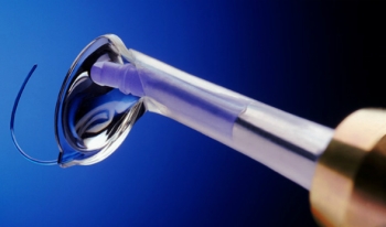
The operation is called phacoemulsification. Using a microsurgical instrument, an incision no larger than 2 mm is made.
Under the action of ultrasound, the substance turns into an emulsion and is removed from the eye through tube systems.
The operation is successful in most cases, but possible complications:
The main drawback of the operation is that as a result of the removal of the lens, the eye loses its ability to accommodate, it is not capable of focusing the image far and near.
If the operation was performed on both eyes, then multifocal glasses are used to achieve image focus on the retinal area.
They have thick lenses and promote distance, near and medium vision. Bifocal glasses are also used, but unlike the previous ones, they provide either distance or near vision.
If the cataract was removed in only one eye the use of contact lenses is advisable. Since the eye of children is constantly growing, then the lenses after a while need to be changed and selected others in size.
Parents should closely monitor the use of contact lenses by children, as the risk of infection is high.
After the operation, it is forbidden to rub your eyes for several days, you can not swim in the pools. Eye drops may be used to moisturize and prevent infection.
Intraocular lens implantation
The ideal way to restore vision is an operation to implant an artificial intraocular lens.
The eye begins to fully function, which manifests itself in the form of image focusing - both far and near.
The intraocular lens implantation operation is also carried out simultaneously and can be combined with cataract removal. Combination is possible in children older than 5-6 years and in adults.
The operation technique is a seamless method. An incision no larger than 2 mm is made, and an intraocular lens is inserted using a microsurgical instrument.
The peculiarity of this lens is its small size (otherwise it simply would not fit into the cut). When placed between the pupil and the vitreous body, the lens expands.
Usually implantation of such a lens is carried out for children not younger than 5 years.
Since the organ of vision in childhood is in a state of constant development, then full recovery of vision should be expected by adolescence
If the operation is late, then amblyopia may develop.. In the preoperative period, due to clouding of the lens, the eye develops incorrectly and "gets used" not to focus a clear image.
In the future after the operation, despite the absence of turbidity, the eye also does not focus the image. This phenomenon is called "lazy eye", or amblyopia.
This condition is difficult to deal with, so it is desirable to prevent it.
Amblyopia is treated with corrective glasses. The second method is eye activation. To do this, a healthy eye is covered with a bandage, and the patient begins to focus images on the retina.
The longer the patient wears the bandage, the better his vision becomes. There are cases when the sharpness was restored to 100%.
The effectiveness of treatment largely depends on the timing of detection. With early detection and further treatment, it is possible to restore vision. Cataract is successfully treated in our country.
The importance of preventing cataracts in children. Excessive strain on vision should be avoided, injury should be avoided and hygiene requirements should be observed.
In contact with
Cataract in children- symptoms and treatment
What is a cataract in children? We will analyze the causes of occurrence, diagnosis and methods of treatment in the article of Dr. Elmanov I. V., an ophthalmologist-surgeon with an experience of 12 years.
Definition of disease. Causes of the disease
Cataract- clouding of the lens, leading to the development of visual deprivation, that is, the deprivation of normal stimulation of the sensory system.
The receptor apparatus of the retina stops working, because due to the cloudy lens, the light does not reach the cones, which convert light stimuli into nerve impulses.

The frequency of occurrence of salivation is 1:10,000 newborns. Bilateral cataracts in children are more common than unilateral ones.
All children's cataracts can be divided into those associated with hereditary anomalies and non-hereditary.
hereditary cataracts:
Non-hereditary cataracts(acquired in utero or in early childhood):
If you experience similar symptoms, consult your doctor. Do not self-medicate - it is dangerous for your health!
Symptoms of cataract in children
One of the early symptoms of congenital cataract is the absence of a red reflex during direct ophthalmoscopy.

Congenital cataract in rare cases is an isolated lesion of the lens. More often it is combined with other eye pathologies: amblyopia, nystagmus, microphthalmos, microcornea and other anomalies of the cornea, as well as the vitreous body, choroid, retina and optic nerve.
Changes in the retina and optic nerve of various nature and severity are the main causes of low visual acuity after extraction of congenital cataract (removal of the cloudy lens). The most common is a combined lesion of the retina and optic nerve. Disorders of the optic nerve suggest its partial atrophy and developmental anomalies (reduction in size, change in the shape of the disc, etc.). On the part of the retina, macular hypoplasia, myelin fibers, central and peripheral dystrophy, and “old” chorioretinal foci are revealed.
![]()
The pathogenesis of cataracts in children
In order to understand the morphology of childhood cataract, it is necessary to understand how the lens develops during embryogenesis (development of the embryo).

The rudiments of the eye appear in the embryo on the 22nd day of intrauterine development. In the process of organogenesis (4-8 weeks of development), an eye bubble appears, which invaginates, turning into an eye cup. The interaction of the eyecup and ectoderm stimulates the development of the lens placode. By the fifth week of intrauterine development, separating from the surface of the ectoderm, a lens vesicle is formed. At the eighth week of development, organelles disappear in the primary lens fibers. Equatorial epithelial cells begin to divide, and new cells, pushing back, lengthen and become secondary lens fibers. At the points of contact of the ends of the secondary lens fibers in the anterior and posterior poles of the lens, linear "seams" are formed. The front seams are U-shaped and the back seams are ʎ.

The final differentiation of fibrillar cells is accompanied by the synthesis and dense packing of crystalline proteins, which create a transparent light-refracting medium. The transparency of the lens is also provided by the programmed disappearance of organelles. At birth, the lens is spherical. Its diameter is 6 mm and in the process of growth of the child increases to 9-10 mm.

An important structural formation associated with the development of congenital cataracts is the pupillary membrane. It forms as a temporary capillary network. This structure becomes most pronounced at 12-13 weeks of gestation. It nourishes the anterior surface of the lens and then must regress.

The stage of regression of the pupillary membrane is an informative marker of the gestational age of preterm infants. At 27-28 weeks, the membrane still completely covers the pupil. At 35-36 weeks of gestation, the pupillary membrane should disappear completely.
The cause of posterior capsular cataract (retrolental plaque) can also be a persistent fetal vitreous vasculature. The hyaloid artery (see figure above) should be completely resorbed (dissolved) by the time the baby is born.
The morphology (structure) of a cataract reflects the age of its occurrence and determines the prognosis for vision. Also, it can sometimes be used to judge the etiology of cataracts. The morphology of childhood cataract is always determined by the anatomy of the lens, its embryology, the duration and nature of the pathological effect that caused it. Acquired cataracts are characterized by a better prognosis for vision than congenital ones.
Some types of congenital cataracts are associated with other eye anomalies. for example microphthalmos often accompanied by nuclear cataract. Autosomal dominant anterior polar cataracts are often associated with Cornea guttata(drip cornea). Wedge-shaped cataracts are characteristic of stickler syndrome.
Classification and stages of cataract development in children
Cataracts in children can be classified into:
By morphology:

According to the degree of cloudiness:
According to the symmetry of the process:
Complications of cataract in children
If during the removal of a cataract, vision in adults can be fully restored, then the child is initially born with an immature visual system, which is formed up to 8-10 years.
Cataract is a serious obstacle to the normal development of the eye, as it causes sensory deprivation and can lead to the development of persistent obstructive amblyopia, strabismus, nystagmus, and the formation of incorrect fixation.
Diagnosis of cataract in children
Anamnesis
Includes family history of cataracts in other children (examination of both parents and siblings), corticosteroid use, radiation therapy, and eye trauma. An examination by related specialists with clarification of concomitant pathology is mandatory. It is important to clarify with parents the age when the child was first diagnosed with strabismus, leukocoria, nystagmus, and abnormal visual behavior. It can be informative to analyze photographs taken with a flash when the child's gaze is directed slightly eccentrically.
Eye status
Visometry (visual acuity assessment) can be difficult due to age, although its determination is possible by optokinetic nystagmus or using special optotypes (for example, Lea optotypes). Indirectly, visual acuity can be judged by the presence or absence of fixation and tracking of objects.

Indirect ophthalmoscopy allows you to establish the localization of lens opacities and examine the fundus.
ultrasound biometrics necessary for diffuse clouding of the lens, when the deep-lying media of the eye are inaccessible to inspection using the previous method.
Biomicroscopy allows to accurately assess the morphology of the cataract, but is not feasible in young children. Most often it is performed intraoperatively. It is difficult to seat a child behind a slit lamp, but it is necessary to examine his parents. This may reveal asymptomatic cataracts. The study also helps to establish the hereditary nature of the disease.
Tonometry to exclude concomitant pathology - congenital glaucoma.
Assessment of the functional state of the visual analyzer(ERG - electroretinography, VEP - visually evoked potentials of the cerebral cortex) allows you to determine the prognosis for vision after cataract extraction.
Laboratory research
Laboratory studies are less indicated in the case of unilateral cataract. Such a study is more informative in the presence of bilateral cataracts, as it indicates the systemic nature of the disease.
These days, many parents deliberately refuse to vaccinate. Therefore, it is necessary to carefully collect an anamnesis, and if a child has a congenital bilateral cataract, it is necessary to determine the titer of antibodies to the IgM rubella virus.
Male infants with cataracts, hypotension, slow weight gain, growth retardation should be screened for Lowe's syndrome. Enzyme analysis (inositol polyphosphate 5‑phosphatase OCRL‑1 in skin fibroblast culture) and an OCRL‑1 test are required to verify the diagnosis.
If you suspect galactosemia should be screened for this disease. Usually it passes in the hospital, but sometimes it is false negative. More than 20 types of mutations are known to cause autosomal dominant cataracts. Most mutations occur in the crystallin and connexin genes.
In general, it can be said that specific tests for suspected congenital syndrome or suspected metabolic disorders are prescribed by a pediatrician or geneticist.
Treatment of cataracts in children
The decision to surgically treat a partially cloudy lens is made only after an assessment of the morphology of the cataract and the visual behavior of the child. The prognosis for vision correlates more with the density than with the size of the opacity. Nuclear cataracts are characterized by the worst prognosis for vision. If it is impossible to distinguish large blood vessels of the fundus through the central zone of the lens, severe visual deprivation should be expected.
Surgery
Surgical treatment of cataracts that threaten the development of visual deprivation should be carried out in infancy. But most surgeons prefer to wait until the baby is 4 weeks old, as there is evidence that cataract surgery before the age of 1 month is associated with a high risk of developing glaucoma. In infants, operations are performed simultaneously on both eyes, especially in children with comorbidities that increase the risk of general anesthesia. But if surgical treatment is delayed, the interval between operations should be short to avoid the risk of developing unilateral amblyopia. The optimal age for surgical treatment of congenital cataract is 3‑6 months.
Lenectomy and anterior vitrectomy in infants are performed on a closed eyeball. Anterior vitrectomy and posterior capsulorhexis can significantly reduce the risk of reoperations. There is no need for cataract phacoemulsification in children; phacoaspiration is performed.

The optimal method for correcting aphakia is the primary implantation of an intraocular lens (IOL). But there are cases when implantation is delayed.
Postoperative management of patients
The main problem of postoperative management of such children is the optical correction of postoperative aphakia (lack of the lens) and the treatment of concomitant amblyopia (“lazy eye”).
Optical stage of treatment
Correction with glasses or contact lenses. Glasses are the simplest and most affordable means of correction. When assigning points, you must follow certain rules:

Aphakic glasses are prescribed if a decision has been made on delayed implantation. They have optical and cosmetic flaws. For delayed IOL implantation, contact lenses are the optimal method of correction.
Pleoptic stage of treatment
A complex of therapeutic and training measures aimed at improving visual acuity. The purpose of pleoptics is to create a functional equality of the eyes, eliminate amblyopia, and correct incorrect visual fixation.
The main component of pleoptic treatment is occlusion. Occlusion can be:
Auxiliary methods of pleoptic treatment:
Orthoptic treatment
It is aimed at restoring normal retinocortical correspondence, that is, at developing normal binocular vision. This type of treatment is possible only with functional equality of the eyes and central fixation.
After the surgical stage of treatment, the child should be under dispensary observation until the age of 12-14. During preventive examinations, refraction is specified, the optical correction necessary at this stage is repeated, courses of pleoptic treatment are repeated, complications after the surgical stage are detected and prevented in a timely manner.
Forecast. Prevention
The results of treatment of children with congenital cataract depend on its early detection, timely (according to indications) surgical treatment, and the presence of concomitant pathology.
Continuity in the work of obstetrician-gynecologists of antenatal clinics, neonatologists of maternity hospitals, pediatricians, oculists of children's clinics and ophthalmologists of specialized medical institutions contributes to the early diagnosis of congenital cataracts and predicting the progression of lens opacities in dynamics.
Timely detection of congenital cataracts in high-risk groups (aggravated hereditary history and complicated course of pregnancy) requires a thorough ophthalmological examination of newborns from the first days of their life - ophthalmoscopy, skiascopy, biomicroscopy under conditions of drug-induced mydriasis (pupil dilation) with an emphasis on the state of the lens. If a pathology of the lens of the child is detected, it is necessary to refer the child to a hospital for a more detailed examination under anesthesia (biomicroscopy, ultrasound and electrophysiological studies) and determine further treatment tactics.
Pronounced changes in the lens, causing visual deprivation, require early surgical intervention. If the lens opacities are insignificant (partial forms of congenital cataract) and do not require early cataract extraction, then an ophthalmologist's dispensary observation is recommended:
According to the indications, in order to prevent and treat amblyopia, correction of refractive errors, pleoptic treatment and medical dilation of the pupil are carried out during this period.
The congenital form is a special type of cataract in a child. The cause of the disease in 30% of cases is due to the impact of any toxin or infection on the fetus in early pregnancy. Congenital cataract in children also occurs with Down syndrome. Scientists have identified a hereditary predisposition to such visual impairment. After delivery in the hospital, each newborn is examined to rule out cataracts.
Cloudiness in the pupil is one of the main signs of cataracts. Pathology is unilateral and bilateral. In addition, there is a change in the shape and size of the lens. Often, along with cataracts in children, doctors diagnose several more anomalies of the organ of vision. It can be strabismus, nystagmus, microophthalmos.
Opacification of the lens looks like a dot, a disk or becomes diffuse. With a nuclear cataract, the pathological site is located in the center of the eye. In the case of the polar variety of the disease, the lens capsule is mainly affected. There may be foci of clouding of the substance at its poles. If the lens is completely cloudy, then the baby is born blind.
The choice of treatment method depends on the shape and size of the cataract. Therapy can be medical or surgical. Occasionally, spontaneous resorption of congenital cataracts occurs with the formation of a film.
Why does the pupil of the eye become white?
Health problems in children and adults are due to a large group of reasons. Experts name among the negative factors affecting the organ of vision, poor ecology, water pollution, infections, nutritional errors. In addition, pregnant women are careless about their health and the unborn baby.
Causes of cataracts in children (congenital)
Congenital cataract is detected in an infant immediately after birth or during the first days of life. Acquired is considered such a form of this disease, in which clouding of the lens occurs at the age of more than two months. The development of congenital cataracts is affected by radiation exposures suffered by a pregnant woman. Especially dangerous in the early stages of gestation are infections - rubella, toxoplasmosis, chicken pox, cytomegalovirus, herpes.

Causes of Acquired Cataract
The maternity hospital provides early diagnosis of congenital cataracts. Then, in the first year of life, an ophthalmologist checks the child's vision during a preventive examination. If a cataract of the eye is suspected, the child is given an instillation of a liquid that dilates the pupil. The doctor then examines the eye with an ophthalmoscope. The absence of a red reflex, as well as clouding of the lens, makes it possible to diagnose a cataract.
Symptoms and treatment of the disease
With a cataract, the pupil becomes cloudy or white, the rays of light almost do not penetrate the lens. Vision deteriorates significantly, the child sees objects unclearly, colors fade for him. Cataract is a serious pathology of the eye that requires immediate treatment. The choice of therapeutic method is influenced by the defeat of one or both pupils, the localization and degree of clouding of the lens.


How to recognize a cataract in a child:
You can suspect a cataract in an infant if the baby in the crib or stroller always follows the bright rattles, tilting his head to one side. Clouding of the lens in children older than 6 years leads to learning problems. The visual system continues to develop until the age of 18, so the treatment of cataracts in children under the age of majority in most cases is carried out with medications. Apply eye drops "Taufon", "Taurine" or Quinax.

The ophthalmologist prescribes the drug "Taufon" to the child, although the instructions indicate its use from the age of 18. The fact is that drops under this trade name have not been studied, although the active substance taurine has been well studied. It is an amino acid that has anti-cataract and metabolic effects. The compound normalizes the functions of cell membranes, stops degenerative changes in the tissues of the eye.
Folk remedies
Alternative medicine prescriptions are also used to treat after consultation with the doctor. The most effective folk remedies are infusions of dill seeds, calendula flowers. The solution is prepared in the standard way, then cotton pads are moistened and applied as a compress or the eyes are washed. It is also recommended to drink tea with calendula flowers - an anti-inflammatory and antioxidant agent.

Vegetable juices improve the work of the visual system - from carrots, parsley and spinach. Red and green vegetables are rich in vitamins A, B 1, B 2, B 12, E, C, flavonoids. Back in 1998, data were published in the English edition of the Ophthalmology journal on the beneficial effect of multivitamins on reducing the incidence of cataracts.
Surgery
If a congenital cataract interferes with the development of the organ of vision, then it is removed at the age of 3–17 weeks. The operation before the child reaches 2 months will avoid further deterioration and loss of vision. The surgery is performed under general anesthesia. The affected lens masses are removed with an irrigation-aspiration device through a tiny incision.
After the operation on the lens, the doctor prescribes glasses to the child, but not immediately. Various complications are possible - increased intraocular pressure, secondary cataract. The resulting inflammatory changes and intraocular pressure are treated with medication. Secondary cataract is removed with a laser. Babies older than 1-4 years can be implanted lenses, artificial lenses.
What is dangerous clouding of the lens in congenital cataracts in children updated: September 19, 2016 by: admin
- partial or complete clouding of the lens of the eye, developing in utero. It manifests itself to varying degrees from the moment the child is born: from a barely noticeable whitish spot to a completely affected lens. Congenital cataract is characterized by a decrease in vision or its complete loss, and strabismus and nystagmus are also noted in children. Primary diagnosis is carried out prenatally; after birth, the diagnosis is confirmed by ophthalmoscopy and slit biomicroscopy. Surgical treatment is shown; lensvitrectomy is performed in the first months of life in uncomplicated cases.
ICD-10
Q12.0

General information
Opacification of the lens occurs by one of two mechanisms. Firstly, initially incorrect laying of the organ of vision. It is characteristic of intrauterine infections in the early stages of pregnancy, chromosomal pathologies, and in general any teratogenic effect if it occurs in the first weeks of pregnancy, when the organ system of vision is formed in the fetus. The second mechanism is the defeat of the already formed lens. It often occurs with metabolic disorders (galactosemia, diabetes mellitus, etc.), exposure to external damaging factors during pregnancy (second or third trimester). In any case, the structure of the lens protein changes, due to which it gradually becomes hydrated and then loses its transparency, as a result of which a congenital cataract develops.
Classification of congenital cataract
The disease is divided into types depending on the location of the turbidity zone and its vastness. There are the following types of cataracts: capsular, polar (anterior and posterior), layered (membraneous), nuclear, complete.
Capsular congenital cataract is a decrease in the transparency of the anterior or posterior lens capsule. The lens itself is not affected. Visual impairment is often minor, but blindness also occurs if the damage to the capsule is extensive, or both the anterior and posterior capsules are affected at the same time.
With a polar cataract, changes affect the anterior or posterior surface of the lens. The capsule is most often also involved in the process. This species is characterized by bilateral lesions. The degree of visual impairment varies greatly.
A stratified cataract is a clouding of one or more of the central layers of the lens. The most common type of congenital cataract is usually bilateral. Vision is usually significantly reduced.
Nuclear cataract is called clouding of the central part of the lens - its nucleus. This variety occurs in all hereditary causes of the disease. The lesion is bilateral, vision is reduced up to complete blindness.
Complete congenital cataract is characterized by clouding of the entire lens. The degree of clouding varies, but more often this form of the disease completely deprives the child of vision. The defeat is bilateral.
By origin, congenital cataract is divided into hereditary and intrauterine. The first of them is transmitted to the child from one of the parents, the second develops in the fetus directly during pregnancy. Atypical (polymorphic) is considered a cataract of complex shape. There are unilateral and bilateral cataracts, and the disease is also classified according to the degree of visual impairment (I-III degrees are distinguished). Some classifications separately designate a complicated form of cataract, but this can be called any clouding of the lens, accompanied by diseases of other organs.
Symptoms of congenital cataract
The main symptom is clouding of the lens of one degree or another. In the clinical picture, it may appear as a noticeable white spot against the background of the iris, but more often there are cases of congenital cataracts when this symptom is absent. With a unilateral lesion, strabismus is noted, as a rule, converging. Sometimes, instead of it, pathological rhythmic trembling of the eyeball is found. Almost all children with bilateral congenital cataracts have nystagmus. At about two months of age, healthy children can already follow the object with their eyes, but this does not happen when they become ill, or the baby always turns towards the object only with his healthy eye.
Diagnosis of congenital cataract
Primary diagnosis is carried out during the planned ultrasound screening of pregnant women. Already in the second trimester, the lens is visualized on ultrasound as a dark spot (normal). There are cases when the second ultrasound still cannot reliably exclude or confirm the diagnosis, and then this can be done in the third trimester. It is important to understand that at this stage the diagnosis cannot be 100% confirmed, but the disease can be suspected, and statistics show that the method is highly reliable.
After the birth of a child, the pediatrician will be able to notice only an intense clouding of the lens of the central localization. Most often, a cataract cannot be diagnosed by physical examination. An examination by a pediatric ophthalmologist is mandatory for all newborns. The specialist will be able to suspect and confirm the diagnosis of congenital cataract, noticing even a slight violation of the passage of light through the lens. Also, the doctor will detect strabismus and nystagmus. Since congenital cataract accompanies many intrauterine infections, metabolic disorders, chromosomal pathologies, when diagnosing these diseases, the child will be examined to exclude eye defects.
For the diagnosis of congenital cataract, the following instrumental methods are used: ophthalmoscopy, slit biomicroscopy, ultrasound of the eyeball. All of them make it possible to verify the change in the transparency of the lens and to exclude similar diseases in the clinic. In particular, retinopathy in children is also characterized by visual impairment and strabismus, however, in this case, the cause is damage to the retina, and examination with an ophthalmoscope will allow this to be established. Tumors of the outer part of the eye can significantly reduce vision, as can congenital cataracts. Visual inspection, ophthalmoscopy, ultrasound methods and X-ray diagnostics help to differentiate them.
Treatment of congenital cataract
Conservative methods of treatment are justified only with a slight clouding of the lens. In therapy, cytoprotectors and vitamins are used. However, most often, congenital cataracts are treated surgically. Surgical removal should be done as early as possible to allow for proper eye development. Cataract surgery - lensvitrectomy - is considered the least traumatic in childhood, therefore it is performed most often. The condition after removal of the lens is called aphakia and requires long-term dynamic observation and vision correction.
Aphakia is corrected with glasses or contact or intraocular lenses. Observation by an ophthalmologist is necessary in order to exclude possible postoperative complications, in particular, glaucoma. A few decades ago, the list of complications was more extensive, but with the introduction of lensvitrectomy, most of them were reduced to a minimum. So, cases of retinal detachment, corneal edema, endophthalmitis and amblyopia are extremely rare.
Prediction and prevention of congenital cataract
Modern methods of surgical treatment provide a favorable prognosis in most cases. Removal of the affected lens at the age of up to six months (better in the first weeks and months) and further vision correction contribute to a good social adaptation of children in adulthood. It should be noted that monocular congenital cataract responds to treatment much worse and currently gives the greatest number of all complications associated with this disease. In addition, cataract is extremely rare in isolation, so the prognosis is also determined by concomitant diseases: infections, metabolic disorders, chromosomal pathologies, etc.
Prevention of congenital cataracts is carried out during pregnancy. It is necessary to exclude the contact of a woman with infectious patients, to minimize the impact of teratogenic factors (alcohol, smoking, radiation methods of diagnosis and therapy, etc.). Women with diabetes should be monitored by an endocrinologist throughout pregnancy. Chromosomal pathologies in most cases are diagnosed before childbirth, and then a woman can already decide to terminate a pregnancy or deliberately carry a child. There is no specific prophylaxis for congenital cataracts.
3985 0
Opacification of the lens- cataracts, accompanied by a decrease in visual acuity from a slight weakening to light perception. There are congenital and acquired - complicated (consecutive) and traumatic.
Congenital cataracts are relatively rare: 5 cases per 100,000 children, according to E. I. Kovalevsky and E. G. Sidorov (1968), however, they account for about 10% of cataracts of various etiologies. Among the causes of blindness in children, congenital cataracts account for 13.2-24.1%, among the causes of low vision - 12.1-13.4%.
Etiology and pathogenesis
Congenital cataracts can be hereditary or arise due to the influence of various teratogenic factors on the lens of the embryo or fetus in the prenatal period.Hereditary cataracts are the result of gene, genomic and chromosomal mutations. More often inherited in an autosomal dominant manner, less often in an autosomal recessive manner. Sex-linked recessive inheritance can be observed, when predominantly men are ill, or dominant, in which women suffer. Hereditary forms account for 25-33% of congenital cataracts, often occurring in several members of the same family.
According to A. V. Khvatova et al. (1985), in 16% of patients with hereditary cataracts, syndromes were observed in which clouding of the lenses was one of the symptoms of genetically determined metabolic disorders. Cataracts can occur with disorders of carbohydrate metabolism. So, congenital cataract is an early sign of galactosemia, in which the synthesis of the enzyme galactose-1-phosphate uridine transferase, contained in erythrocytes and the lens, is impaired.
In addition to cataracts, which usually develop in the first half of life, jaundice, hepatosplenomegaly, cirrhosis of the liver, mental retardation, etc. are observed with this disease. The occurrence of pathological symptoms in galactosemia can be prevented by promptly excluding milk and dairy products from the child's diet. With early implementation of these measures in the initial period of cataract development (in children under 3 months), lens opacity may regress.
With hypoglycemia, cataract occurs, as a rule, at the 2-3rd month of life. Opacification of the lens occurs in 1-4% of patients with diabetes, which usually develops between the ages of 6 and 13 years, but also occurs in young children, even in infants. Cataract in diabetes progresses rapidly, indicating a severe course of the disease. Clouding of the lens, along with clouding of the cornea, damage to the optic nerve, strabismus, is one of the symptoms of mannosidosis, a metabolic disorder associated with a deficiency of active acid mannosidase A and B. The disease is characterized by multiple dysostoses, hearing loss, hepatosplenomegaly, psychomotor retardation, etc.
In cases of violation of calcium metabolism, the so-called tetanic cataract (usually zonular)), tetany, spasmophilia, rickets, renal failure can be observed at the same time. Treatment started at an early stage of the disease (intravenous injections of 5-10% calcium chloride solution, thyroid drugs) can stop the development of lens opacity.
Cataract is one of the symptoms of hereditary changes in the connective tissue and abnormalities of the skeletal system. Together with other ocular and general somatic manifestations, it is observed in Marfan and Marchezani syndromes. Cataract is one of the constant symptoms of congenital chondrodystrophy, characterized by a slowdown in the epiphyseal growth of long bones with normal periosteal growth, which leads to shortening and thickening of the bones.
Clouding of the lens is often observed in Apert's syndrome, the signs of which are a "tower" skull, syndactyly, polydactyly, hypoplasia of the upper jaw, heart disease, mental retardation, flattening of the orbits, exophthalmos, divergent strabismus, nystagmus, damage to the optic nerve (congestive discs, atrophy), colobomas of the choroid, retinal damage. Congenital or developing cataracts in the first year of life are often found in Conradi syndrome, characterized by disproportionate dwarfism, finger anomalies, scoliosis, and damage to the ligamentous apparatus of the joints.
Clouding of the lens in hereditary skin lesions is explained by the fact that it is a derivative of the ectoderm. Cataract develops in Rothmund's syndrome, characterized by dermatosis, hypogenitalism, and congenital ichthyosis, which is a combination of hyperkeratosis with damage to the organ of vision. With Schaefer's syndrome, along with follicular keratosis of the skin, foci of baldness, microcephaly, dwarf growth, hypogenitalism, mental retardation, congenital cataracts are observed.
Opacification of the lens is also characteristic of infantile pigmentary dermatosis Bloch-Sulzberger, in which skin pigmentation, alopecia, anomalies of the teeth, and other eye lesions are noted - atrophy of the optic nerves, strabismus, nystagmus. Congenital cataract, together with microphthalmos, heterochromia, ectopia of the pupil, corneal opacity, color blindness, is observed in constitutional spherical cell hemolytic anemia, the general somatic manifestations of which are hemolytic jaundice, splenomegaly, bone anomalies, impaired pigmentation of the skin, otosclerosis, polydactyly, etc.
Of the chromosomal diseases with damage to the lens, Down's disease is more common than others, which is caused by trisomy of chromosome 21 and is observed mainly in children of elderly mothers. With this disease, severe disorders of physical and mental development are detected.
Characteristic signs of Down's disease are idiocy or imbecility, micro- and brachycephaly, wide sunken bridge of the nose, "geographical" tongue, underdevelopment of the upper jaw, short stature or acromegaly, hypothyroidism, ichthyosis, keratosis, alopecia, hypogenitalism, syndactyly, polydactyly, congenital heart disease and other anomalies. About half of patients die mainly in the first year of life due to the reduced resistance of their body to infections, some live to a ripe old age. Eye manifestations of Down's disease are epicanthus, nystagmus, strabismus, corneal clouding; cataract occurs in 15-50% of patients.
Chromosomal disorders that cause cataracts and other eye abnormalities include: Marinescu-Sjögren syndrome, characterized by congenital cerebellar spinal ataxia, delayed physical and mental development, dwarf growth; Shereshevsky-Turner-Bonnevie-Ulrich syndrome (neck pterygium syndrome); Low's syndrome; Norrie disease.
There are numerous other syndromes, one of the manifestations of which is congenital cataract: Axenfeld, Alport, Cockayne, Rieger, and others. Cataracts have been described in the syndromes of Marten-Albright, Bassen Kornzweig, Knapp-Komrover, Saburo, and others.
Most congenital cataracts occur as a result of intrauterine development disorders due to the influence of various adverse factors (physical, chemical, biological) on the lens of the embryo or fetus, both external and internal. These can be various intoxications (alcohol, ether, some contraceptives and abortifacients, sleeping pills), ionizing radiation, hypovitaminosis (deficiency of vitamins A, E, folic and pantothenic acids), Rh incompatibility of the mother and fetus, oxygen starvation of the fetus due to circulatory disorders and others
The cause of the development of congenital cataracts, as well as other anomalies, can be various diseases of the mother during pregnancy: cardiovascular (for example, rheumatic heart disease with circulatory failure), endocrine disorders, etc. Of particular importance are infectious diseases caused by bacteria, protozoa (toxoplasma), viruses. Most viruses are able to cross the placental barrier and infect the fetus and fetus, causing the development of cataracts and other anomalies that can occur with rubella, cytomegaly, chickenpox, herpes, and influenza. Infectious diseases of the mother during pregnancy, in particular rubella, often occur without characteristic symptoms, which further complicates the etiological diagnosis of cataracts.
Depending on the time of exposure to the teratogenic factor, the nature and degree of its influence on the organ of vision of the embryo and fetus, various forms of cataracts and concomitant changes in the organ of vision occur. The most dangerous period of exposure to teratogenic factors on the organ of vision is the 3-7th week of pregnancy.
Clinical features of congenital cataracts. The clinical picture of congenital cataracts is characterized by significant variability depending on the intensity, form and localization of lens opacity, as well as the presence or absence of other eye defects (Table 24, Fig. 105).
Rice. 105. Scheme of various forms of congenital cataracts: 1 - complete; 2 - semi-absorbed; 3 - central; 4 - zonular; 5 - spindle-shaped; 6 - front and back polar; 7 - pyramidal
Table 24. Classification of congenital cataracts

Complete (diffuse) cataract is characterized by total clouding of the lens, the shape and dimensions of which are preserved (Fig. 106). The reflex from the fundus is absent in both narrow and dilated pupils. Vision is reduced to light perception. A variety of complete cataract is the so-called milky cataract, the peculiarity of which is the liquefaction of the clouded lens substance.

Zonular cataract is a partial clouding of the lens in the form of separate layers (or layers) located between the embryonic nucleus and cortical layers (Fig. 107), the rest of the lens is transparent, sometimes two or three layers of clouding are observed, separated by a transparent substance of the lens. Opacities have the form of a disk of various diameters (usually 3-9 mm) with clear, even edges. At the equator, the disk, as a rule, is surrounded by the so-called riders - opacities of the perinuclear zone, which have the shape of a loop or hook. Their number is different.

Rice. 107. Zonular cataract
In lateral illumination, the layered cataract looks like a gray disc. In the study in transmitted light against the background of a pink-red reflex from the fundus of the eye, a disk of dark clouding is visible. With its relatively small size, it is possible to examine the fundus of the eye and even study refraction through the transparent periphery of the lens. Visual acuity in zonular cataracts, depending on the size of the disk and the intensity of clouding, as well as the severity of concomitant changes in the organ of vision, ranges from several hundredths to several tenths, reaching 0.3-0.5 in some cases. As the pupil dilates, visual acuity usually increases.
Nuclear (central) cataract is characterized by clouding of the embryonic nucleus of the lens, the transparency of the remaining parts of which is preserved. In lateral illumination, the cataract looks like a small gray disk in the center of the lens; in transmitted light, against the background of a pink-red reflex, a central dark disk is visible from the fundus. Visual acuity is significantly reduced, with pupil dilation it can increase.
A semi-resolved cataract is manifested in a decrease in the volume of the lens due to partial spontaneous resorption of it, which occurs in the prenatal or postnatal period. A semi-dissolved cataract is more often formed from a complete one and is a diffuse opacification of a flattened lens (its anteroposterior size is reduced with a normal diameter), accompanied by a decrease in vision to light perception.
In cases of formation of a semi-absorbed cataract from a zonular cataract, destruction of the transparent zone is observed; at the same time, uniform vision can be preserved, which decreases as the process of lens resorption progresses.
A membranous cataract is a flat membrane consisting of a clouded anterior capsule (often with fibrous deposits on it) and a posterior lens capsule, between which there may be remnants of the lens substance. A cataract is formed as a result of intrauterine or postnatal resorption of the lens substance or is an anomaly in the development of the lens.
Filmy cataracts can be of different density, often of uneven thickness. The reflex from the fundus is often absent, in some cases it is slightly pink (through thinned areas of the cataract). Vision is usually reduced to light perception or a few hundredths.
Atypical (polymorphic) cataract characterized by partial clouding of the lens of various shapes, localizations and sizes (Fig. 108). Depending on this, vision is reduced to varying degrees. Cataracts can be either lenticular or capsulo-lenticular.

Rice. 108. Atypical (polymorphic) cataracts (a, b)
polar cataract- limited capsulolenticular opacification at the anterior (anterior polar cataract) or posterior (posterior polar cataract) pole of the lens. This localization of opacification is explained by the fact that the poles at which the sutures of the lens converge are the places of least resistance. The formation of anterior polar cataract is associated with the presence of remnants of the pupillary membrane and a violation of the process of detachment of the lens vesicle from the outer ectoderm in the early period of embryo development.
In the pathogenesis of posterior polar cataract, the weakness of the lens capsule at the posterior pole is given importance, in connection with which its rupture may occur, accompanied by protrusion and limited clouding of the lens substance, as well as the penetration of mesenchymal elements through the rupture. Capsule ruptures may occur as a result of posterior traction during regression of the vitreous artery.
Polar cataract (anterior or posterior) is a round, dense or stratified whitish-gray opacification of the capsule and adjacent layers of the lens. In transmitted light, against the background of a pink-red reflex from the fundus, a rounded, dark, usually slight clouding is visible.
Posterior polar cataract should be differentiated from a false cataract, which is the remnants of a reduced vitreous artery and vitreous (cloquets) canal. False cataracts are usually medial to the posterior pole and the opacities are located on the posterior capsule on the outside of the lens, while in polar cataracts they fit into the lens section.
Polar cataracts either do not affect visual acuity, or do not significantly reduce it. With unilateral congenital cataracts, obscurative amblyopia may develop, in these cases visual acuity does not correspond to the amount of clouding.
A variety of anterior polar cataract is: pyramidal cataract, in which homogeneous or layered opacity has the shape of a cone facing the anterior chamber.
Anterior and posterior capsulolenticular opacities can be connected by spindle-shaped opacities, forming a spindle-shaped cataract, in which visual acuity is reduced more significantly than in polar cataracts, but, as a rule, remains at a fairly high level.
There are a large number of other forms of congenital cataracts with partial, limited opacities, which usually do not affect visual acuity (or slightly reduce it), they are usually detected during preventive examinations. These cataracts include coronary (coronary), characterized by numerous limited opacities, greenish-bluish in color, which are located in the middle and deep layers along the periphery of the lens like a wreath. With anterior axial embryonic cataract, there is clouding of the lens near its anteroposterior axis in the region of the anterior epsilon-shaped suture of the embryonic nucleus.
Star-shaped suture cataract is characterized by clouding in the region of the anterior or posterior sutures of the lens. Multiple very small grayish-blue opacities, diffusely scattered in the region of the embryonic nucleus, are observed with punctate cataracts. For nuclear powdery cataracts, diffuse opacification of the embryonic nucleus, consisting of dust-like elements, is characteristic.
Congenital cataract relatively rarely represents an isolated lesion of the lens, it is often combined with other pathological changes in the organ of vision, as well as other organs and systems of the child's body. Various defects of the organ of vision are observed in 36.8-77.3% of children with congenital cataracts: strabismus, nystagmus, microphthalmos, microcornea, pathology of the cornea, vitreous body, retina and optic nerve.
Strabismus is observed in a large number of children (up to 83%) with congenital cataracts. In some cases, it can be a complication of cataracts, the result of a sharp decrease in vision and deep disturbances in the sensory-motor connections of the visual analyzer. In others, strabismus is a congenital pathology associated with cataracts. Strabismus in congenital cataracts is more often converging, alternating, mostly permanent. The angle of deviation of the eye varies from 5 to 60%, more often it is 15-20°.
nystagmus, observed in children with congenital cataracts, may be congenital and acquired, associated with a sharp decrease in vision. Pendulum-shaped and mixed nystagmus is more common, less often jerk-like, it is predominantly horizontal, small-caliber. Most often, nystagmus is observed with complete, membranous cataracts, combined with other defects of the organ of vision, such as microphthalmos, microcornea, etc.
microphthalmos, characterized by a decrease in the size of the eyeball and functional inferiority of the organ of vision, is observed in children with congenital cataracts. The causes of congenital microphthalmos can be hereditary and genetic factors, as well as intrauterine inflammatory and degenerative processes that retard the growth of the eyeball. The degree of reduction of the eye is different. With microphthalmos, other malformations of the eye can be observed: colobomas of the vascular tract and optic nerve, macular aplasia, etc.
Microcornea or small cornea, in most cases it is noted with microphthalmos, but it can also occur on its own. With congenital cataracts, other anomalies of the cornea can be observed: its opacities, dermoid cysts, megalocornea.
Congenital cataracts are often combined with various changes in the vitreous body, in particular with its opacities of varying severity - from small to coarse fibrous. A frequent finding are the remains of an incompletely reduced vitreous artery.
Anomalies of the choroid in congenital cataracts are colobomas of the iris and choroid, polycoria, pupil displacement. Hypoplasia and aplasia of the dilator, hypoplasia of the iris are often observed. Less common is dysgenesis of the iris and cornea, the manifestations of which are posterior embryotoxon, hyaline films and thickenings on the posterior surface of the cornea, and peripheral anterior adhesions.
Fundus changes detected after removal of congenital cataracts include partial atrophy of the optic nerve, myelin fibers, chorioretinal lesions, macular hypoplasia, retinal dystrophy, pigment redistribution, etc.
Obscurative amblyopia, or more precisely, underdevelopment of the visual analyzer as a result of the absence of a light stimulus of the retina, is a severe complication of congenital lens opacities, which is the most common cause of low visual acuity after cataract removal and discrepancy between functional results and the optical effect of the operation.
The most severe obscurative amblyopia develops with complete (diffuse) lens opacities. In order to prevent damage to the visual analyzer, a method has been proposed that consists in permanent dilation of the pupils with the help of mydriatics and subsequent irritation of the eyes with light stimuli. These manipulations should be performed in the first 6 months of life, if by this time an operation (cataract extraction) has not been performed.
Treatment
Surgical treatment of congenital cataracts. Most authors recommend removing congenital cataracts with visual acuity below 0.3. After cataract extraction, aphakia correction, pleoptic and, according to indications, orthotopic treatment, measures aimed at eliminating strabismus, and treatment of nystagmus are carried out.The issue of the timing of removal of congenital cataract is decided individually based on the clinical form of cataract, residual visual acuity, cataract etiology, and the general condition of the child. Due to the risk of obscurative (deprivation) amblyopia with the long-term existence of congenital cataracts, as well as the need to increase visual acuity in order for the child to develop, it is advisable to perform the operation at an early date.
According to the majority of authors who studied changes in the visual analyzer under conditions of deprivation, the sensitive period of vision development falls on the period from the 2nd to the 6th month of a child's life. In this regard, the specified age is optimal for the removal of congenital cataracts in children if there are indications for early surgery.
Early surgery is indicated for: 1) complete, semi-absorbed, membranous cataracts; 2) zonular, central, atypical cataracts with a turbidity diameter of more than 3 mm, when retinoscopy is impossible under conditions of mydriasis; 3) the presence of a rigid pupil in all forms of congenital cataracts due to the fact that in these cases it is impossible to achieve mydriasis, which is necessary for preoperative measures to prevent amblyopia and retinal underdevelopment; 4) the appearance of nystagmus in any form of cataract, which indicates a violation of the development of the oculomotor reflex.
Early surgery is not indicated for zonular cataracts with an opacity disk of less than 3 mm, in which retinoscopy can be performed due to the presence in these cases, as a rule, of relatively high residual visual acuity and the absence of the risk of developing severe amblyopia. In such patients, surgery may be performed at a later date.
It should be borne in mind that with congenital cataracts, the development of which is associated with intrauterine infection (rubella, etc.), complications (iridocyclitis) may occur after an early operation.
Unilateral congenital cataracts should be removed at almost the same time as bilateral ones.
For any form of cataract, surgery should be postponed in cases where there are general contraindications for surgical intervention and anesthesia (acute infections, chronic diseases in the acute and decompensated stages, the presence of foci of focal infection, allergic status, enlarged thymus gland, etc.). The operation in these cases is carried out after treatment with the permission of the relevant specialists. In case of cataracts, the origin of which is associated with hereditary metabolic disorders (galactosemia, homocystinuria, etc.), it is necessary to carry out corrective treatment before surgery.
Contraindications for removal of congenital cataracts there may be extensive inoperable retinal detachment, gross fibrosis of the vitreous body in combination with retinal damage, etc. However, indications and contraindications for surgery in these cases are established individually based on the results of a clinical and functional study of the organ of vision.
Due to age-related characteristics (strength of the ligamentous apparatus of the lens), an extracapsular method is used to remove cataracts in children. Intracapsular extraction in childhood is not used due to the high trauma of the operation and the possibility of developing severe complications due to the strength of the fibers of the ciliary girdle and hyaloid-capsular ligament, the close connection between the posterior lens capsule and the anterior limiting membrane of the vitreous body. The use of zonulolytic agents to dissolve the fibers of the ciliary girdle in children is not recommended.
One of the oldest treatments for congenital cataracts- discission. The principle of the operation is to cut the anterior lens capsule with a dissection needle, as a result of which the lens substance enters the anterior chamber, swells and dissolves. The disadvantages of the method are the frequent complications associated with leaving the lens substance in the eye: the formation of a secondary cataract, secondary glaucoma, phacogenic iridocyclitis, as well as the slow resorption of the cataract, and therefore the visual effect of the operation appears after a few months, during which the conditions for increasing the obscuration remain. amblyopia.
Due to these shortcomings, discission is not currently used for soft cataracts. A two-stage operation, in which discission is first performed, and after 2-3 weeks, when the lens substance swells, it is removed or aspirated, is also not without these shortcomings, therefore it cannot be recommended.
In case of zonular cataracts with a small (not more than 5.0-5.5 mm) opacity disk and a significant increase in visual acuity with pupil dilation by mydriatic means, an optical iridectomy is performed, the advantage of which is the preservation of accommodation. However, the method has a number of significant drawbacks: vision is not fully restored due to the preservation of an optical obstacle in the form of a central clouding of the lens, the shape of the pupil changes, so it is not advisable to use it.
In congenital cataracts, various modifications of extracapsular (linear) extraction are used. The principle of the operation is extracapsular cataract removal through a small (2.5-3.0 mm) corneal incision.
In the surgical treatment of congenital cataracts, it is widely used aspiration method. The principle of the operation is to aspirate the lens substance. The advantage of aspiration is the ability to remove congenital cataracts through a small incision (1.5-2.0 mm), which ensures the safety of the method. A cystotome introduced into the anterior chamber through an incision in the cornea, limbus, or limbus under the conjunctival flap opens the anterior lens capsule, the substance of which is sucked off through a cannula connected to a syringe.
It is advisable to use aspiration-irrigation technique, which provides greater efficiency and less traumatic operation. Thanks to the irrigation of the liquid carried out simultaneously with aspiration (most often isotonic sodium chloride solution), a constant depth of the anterior chamber is maintained, and therefore the possibility of injury to the endothelium of the cornea and iris is excluded.