
The sequence of passage of a sound signal through the organ of hearing. Structure and functions of the outer and middle ear
The Hearing and Balance Organ is the peripheral part of the Gravity, Balance and Hearing Analyzer. It is located within one anatomical formation - the labyrinth and consists of the outer, middle and inner ear(Fig. 1).
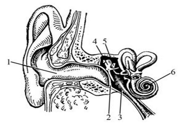
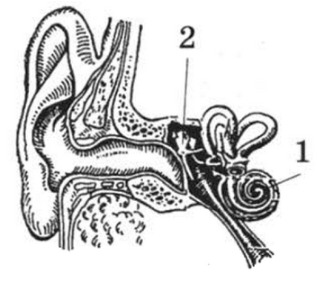
Rice. 1. (diagram): 1 - external auditory canal; 2 - auditory tube; 3 - eardrum; 4 - hammer; 5 - anvil; 6 - snail.
1. Outer ear(auris externa) consists of the auricle (auricula), external auditory canal (meatus acusticus externus), and eardrum(membrana tympanica). The outer ear plays the role of the auditory funnel to capture and conduct sound.
Between the external auditory canal and the tympanic cavity is the eardrum (membrana tympanica). The eardrum is elastic, low-elastic, thin (0.1-0.15 mm thick), and concave inward in the center. The membrane has three layers: dermal, fibrous and mucous. It has a loose part (pars flaccida) - Shrapnel membrane, which does not have a fibrous layer, and a tense part (pars tensa). For practical purposes, the membrane is divided into squares.
2. Middle ear(auris media) consists of the tympanic cavity (cavitas tympani), auditory tube (tuba auditiva) and mastoid cells (cellulae mastoideae). The middle ear is a system of air cavities in the thickness of the petrous part of the temporal bone.
Tympanic cavity has a vertical dimension of 10 mm and a transverse dimension of 5 mm. The tympanic cavity has 6 walls (Fig. 2): lateral - membranous (paries membranaceus), medial - labyrinthine (paries labyrinthicus), anterior - carotid (paries caroticus), posterior - mastoid (paries mastoideus), superior - tegmental (paries tegmentalis) ) and lower - jugular (paries jugularis). Often in top wall there are cracks in which the mucous membrane of the tympanic cavity is adjacent to the dura mater.

Rice. 2. : 1 - paries tegmentalis; 2 - paries mastoideus; 3 - paries jugularis; 4 - paries caroticus; 5 - paries labyrinthicus; 6 - a. carotis interna; 7 - ostium tympanicum tubae auditivae; 8 - canalis facialis; 9 - aditus ad antrum mastoideum; 10 - fenestra vestibuli; 11 - fenestra cochleae; 12 - n. tympanicus; 13 - v. jugularis interna.
The tympanic cavity is divided into three floors; supratympanic recess (recessus epitympanicus), middle (mesotympanicus) and lower - subtympanic recess (recessus hypotympanicus). In the tympanic cavity there are three auditory ossicles: the malleus, the incus and the stapes (Fig. 3), two joints between them: the incus-malleus (art. incudomallcaris) and the incudostapedialis (art. incudostapedialis), and two muscles: the tensor tympani ( m. tensor tympani) and stirrup (m. stapedius).

Rice. 3. : 1 - malleus; 2 - incus; 3 - steps.
Eustachian tube- channel 40 mm long; has a bony part (pars ossea) and a cartilaginous part (pars cartilaginea); connects the nasopharynx and the tympanic cavity with two openings: ostium tympanicum tubae auditivae and ostium pharyngeum tubae auditivae. During swallowing movements, the slit-like lumen of the tube expands and freely passes air into the tympanic cavity.
3. Inner ear(auris interna) has a bony and membranous labyrinth. Part bony labyrinth(labyrinthus osseus) included semicircular canals, vestibule And cochlea canal(Fig. 4).
Membranous labyrinth(labyrinthus membranaceus) has semicircular ducts, little queen, pouch And cochlear duct(Fig. 5). Inside the membranous labyrinth there is endolymph, and outside there is perilymph.

Rice. 4.: 1 - cochlea; 2 - cupula cochleae; 3 - vestibulum; 4 - fenestra vestibuli; 5 - fenestra cochleae; 6 - crus osseum simplex; 7 - crura ossea ampullares; 8 - crus osseum commune; 9 - canalis semicircularis anterior; 10 - canalis semicircularis posterior; 11 - canali semicircularis lateralis.

Rice. 5. : 1 - ductus cochlearis; 2 - sacculus; 3 - utriculus; 4 - ductus semicircularis anterior; 5 - ductus semicircularis posterior; 6 - ductus semicircularis lateralis; 7 - ductus endolymphaticus in aquaeductus vestibuli; 8 - saccus endolymphaticus; 9 - ductus utriculosaccularis; 10 - ductus reuniens; 11 - ductus perilymphaticus in aquaeductus cochleae.
The endolymphatic duct, located in the aqueduct of the vestibule, and the endolymphatic sac, located in the cleft of the dura mater, protect the labyrinth from excessive vibrations.
On a cross section of the bony cochlea, three spaces are visible: one endolymphatic and two perilymphatic (Fig. 6). Because they climb up the coils of the cochlea, they are called staircases. The median staircase (scala media), filled with endolymph, has a triangular outline in cross-section and is called the cochlear duct (ductus cochlearis). The space located above the cochlear duct is called the scala vestibuli; the space located below is the scala tympani.

Rice. 6. : 1 - ductus cochlearis; 2 - scala vestibuli; 3 - modiolus; 4 - ganglion spirale cochleae; 5 - peripheral processes of ganglion spirale cochleae cells; 6 - scala tympani; 7 - bone wall of the cochlear canal; 8 - lamina spiralis ossea; 9 - membrane vestibularis; 10 - organum spirale seu organum Cortii; 11 - membrane basilaris.
Sound path
Sound waves are picked up by the auricle, sent to the external auditory canal, and cause vibrations in the eardrum. The vibrations of the membrane are transmitted by the system of auditory ossicles to the window of the vestibule, then to the perilymph along the scala vestibule to the apex of the cochlea, then through the lucid window, the helicotrema, to the perilymph of the scala tympani and are attenuated, hitting the secondary tympanic membrane in the cochlear window (Fig. 7).

Rice. 7. : 1 - membrana tympanica; 2 - malleus; 3 - incus; 4 - steps; 5 - membrana tympanica secundaria; 6 - scala tympani; 7 - ductus cochlearis; 8 - scala vestibuli.
Through the vestibular membrane of the cochlear duct, vibrations of the perilymph are transmitted to the endolymph and the main membrane of the cochlear duct, on which the receptor of the auditory analyzer, the organ of Corti, is located.
Conducting path of the vestibular analyzer
Receptors of the vestibular analyzer: 1) ampullary scallops (crista ampullaris) - perceive the direction and acceleration of movement; 2) spot of the uterus (macula utriculi) - gravity, position of the head at rest; 3) sac spot (macula sacculi) - vibration receptor.
The bodies of the first neurons are located in the vestibular node, g. vestibulare, which is located at the bottom of the internal auditory canal (Fig. 8). The central processes of the cells of this node form the vestibular root of the eighth nerve, n. vestibularis, and end on the cells of the vestibular nuclei of the eighth nerve - the bodies of the second neurons: upper core- core V.M. Bekhterev (there is an opinion that only this nucleus has a direct connection with the cortex), medial(main) - G.A Schwalbe, lateral- O.F.C. Deiters and lower- Ch.W. Roller. The axons of the cells of the vestibular nuclei form several bundles, which are sent to the spinal cord, to the cerebellum, to the medial and posterior longitudinal fasciculi, as well as to the thalamus.

Rice. 8.: R - receptors - sensitive cells of the ampullary combs and cells of the spots of the utricle and sac, crista ampullaris, macula utriculi et sacculi; I - first neuron - cells of the vestibular node, ganglion vestibulare; II - second neuron - cells of the superior, inferior, medial and lateral vestibular nuclei, n. vestibularis superior, inferior, medialis et lateralis; III - third neuron - lateral nuclei of the thalamus; IV - cortical end of the analyzer - cells of the cortex of the inferior parietal lobule, middle and inferior temporal gyri, Lobulus parietalis inferior, gyrus temporalis medius et inferior; 1 - spinal cord; 2 - bridge; 3 - cerebellum; 4 - midbrain; 5 - thalamus; 6 - internal capsule; 7 - area of the cortex of the inferior parietal lobule and the middle and inferior temporal gyri; 8 - vestibulospinal tract, tractus vestibulospinalis; 9 - motor nucleus cell anterior horn spinal cord; 10 - cerebellar tent nucleus, n. fastigii; 11 - vestibulocerebellar tract, tractus vestibulocerebellaris; 12 - to the medial longitudinal fasciculus, reticular formation and the vegetative center of the medulla oblongata, fasciculus longitudinalis medialis; formatio reticularis, n. dorsalis nervi vagi.
The axons of the cells of the Deiters and Roller nuclei enter the spinal cord, forming the vestibulospinal tract. It ends on the cells of the motor nuclei of the anterior horns of the spinal cord (the bodies of the third neurons).
The axons of the cells of the Deiters, Schwalbe and Bechterew nuclei are sent to the cerebellum, forming the vestibulocerebellar tract. This pathway passes through the inferior cerebellar peduncles and ends at the cells of the cerebellar vermis cortex (the body of the third neuron).
The axons of the cells of the Deiters nucleus are sent to the medial longitudinal fasciculus, which connects the vestibular nuclei with the nuclei of the third, fourth, sixth and eleventh cranial nerves and ensures that the direction of gaze is maintained when the position of the head changes.
From Deiters' nucleus, axons are also sent to the posterior longitudinal fasciculus, which connects the vestibular nuclei with the autonomic nuclei of the third, seventh, ninth and tenth pairs of cranial nerves, which explains autonomic reactions in response to excessive stimulation of the vestibular apparatus.
Nerve impulses to the cortical end of the vestibular analyzer pass as follows. The axons of the cells of the Deiters and Schwalbe nuclei pass to the opposite side as part of the vestibular tract to the bodies of the third neurons - the cells of the lateral nuclei of the thalamus. The processes of these cells pass through the internal capsule into the cortex of the temporal and parietal lobes of the hemisphere.
Conducting path of the auditory analyzer
Receptors that perceive sound stimulation are located in the organ of Corti. It is located in the cochlear duct and is represented by sensory hair cells located on the basement membrane.
The bodies of the first neurons are located in the spiral ganglion (Fig. 9), located in the spiral canal of the cochlea. The central processes of the cells of this node form the cochlear root of the eighth nerve (n. cochlearis) and end on the cells of the ventral and dorsal cochlear nuclei of the eighth nerve (the bodies of the second neurons).

Rice. 9.: R - receptors - sensitive cells of the spiral organ; I - first neuron - cells of the spiral ganglion, ganglion spirale; II - second neuron - anterior and posterior cochlear nuclei, n. cochlearis dorsalis et ventralis; III - third neuron - anterior and posterior nuclei of the trapezoid body, n. dorsalis et ventralis corporis trapezoidei; IV - fourth neuron - cells of the nuclei of the inferior colliculi of the midbrain and medial geniculate body, n. colliculus inferior et corpus geniculatum mediale; V - cortical end of the auditory analyzer - cells of the cortex of the superior temporal gyrus, gyrus temporalis superior; 1 - spinal cord; 2 - bridge; 3 - midbrain; 4 - medial geniculate body; 5 - internal capsule; 6 - section of the cortex of the superior temporal gyrus; 7 - roof-spinal tract; 8 - cells of the motor nucleus of the anterior horn of the spinal cord; 9 - fibers of the lateral loop in the loop triangle.
The axons of the cells of the ventral nucleus are directed to the ventral and dorsal nuclei of the trapezoidal body on their own and the opposite side, and the latter form the trapezoidal body itself. The axons of the cells of the dorsal nucleus pass to the opposite side as part of the medullary striae, and then the trapezoid body to its nuclei. Thus, the bodies of third neurons auditory pathway located in the nuclei of the trapezoid body.
The totality of axons of third neurons is lateral loop(lemniscus lateralis). In the isthmus region, the loop fibers lie superficially in the loop triangle. The fibers of the loop end on the cells of the subcortical centers (the bodies of the fourth neurons): the inferior colliculi of the quadrigeminal and the medial geniculate bodies.
The axons of the cells of the nucleus of the inferior colliculus are directed as part of the roof-spinal tract to the motor nuclei of the spinal cord, carrying out unconditioned reflex motor reactions of the muscles to sudden auditory stimulation.
The axons of the cells of the medial geniculate bodies pass through the posterior leg of the internal capsule into the middle part of the superior temporal gyrus - the cortical end of the auditory analyzer.
There are connections between the cells of the nucleus of the inferior colliculus and the cells of the motor nuclei of the fifth and seventh pairs cranial nuclei, providing regulation of the auditory muscles. In addition, there are connections between the cells of the auditory nuclei with the medial longitudinal fasciculus, which ensure the movement of the head and eyes when searching for a sound source.
Development of the vestibulocochlear organ
1. Development of the inner ear. The rudiment of the membranous labyrinth appears in the 3rd week of intrauterine development through the formation of thickenings of the ectoderm on the sides of the anlage of the posterior medullary vesicle (Fig. 10).

Rice. 10.: A - stage of formation of auditory placodes; B - stage of formation of auditory pits; B - stage of formation of auditory vesicles; I - first visceral arch; II - second visceral arch; 1 - pharyngeal intestine; 2 - medullary plate; 3 - auditory placode; 4 - medullary groove; 5 - auditory fossa; 6 - neural tube; 7 - auditory vesicle; 8 - first gill pouch; 9 - first gill slit; 10 - growth of the auditory vesicle and formation of the endolymphatic duct; 11 - formation of all elements of the membranous labyrinth.
At stage 1 of development, the auditory placode is formed. At stage 2, an auditory fossa is formed from the placode, and at stage 3, an auditory vesicle is formed. Next, the auditory vesicle lengthens, the endolymphatic duct protrudes from it, which pulls the vesicle into 2 parts. The semicircular ducts develop from the upper part of the vesicle, and the cochlear duct develops from the lower part. Receptors for the auditory and vestibular analyzers are formed in the 7th week. The cartilaginous labyrinth develops from the mesenchyme surrounding the membranous labyrinth. It ossifies in the 5th week of intrauterine development.
2. Middle ear development(Fig. 11).
The tympanic cavity and auditory tube develop from the first gill pouch. Here a single tubular-drum canal is formed. The tympanic cavity is formed from the dorsal part of this canal, and the auditory tube is formed from the dorsal part. From the mesenchyme of the first visceral arch the hammer, incus, m. tensor tympani, and the fifth nerve innervating it, from the mesenchyme of the second visceral arch - the stapes, m. stapedius and the seventh nerve that innervates it.

Rice. 11.: A - location of the visceral arches of the human embryo; B - six tubercles of mesenchyme located around the first external gill slit; B - auricle; 1-5 - visceral arches; 6 - first gill slit; 7 - first gill pouch.
3. Development of the outer ear. The auricle and external auditory canal develop as a result of the fusion and transformation of six tubercles of mesenchyme located around the first external branchial cleft. The pit of the first external gill slit deepens, and a tympanic membrane is formed in its depth. Its three layers develop from three germ layers.
Anomalies in the development of the hearing organ
- Deafness can be a consequence of underdevelopment of the auditory ossicles, a violation of the receptor apparatus, as well as a violation of the conductive part of the analyzer or its cortical end.
- Fusion of the auditory ossicles, reducing hearing.
- Anomalies and deformities of the external ear:
- anotia - absence of the auricle,
- buccal auricle,
- fused lobe,
- shell consisting of one lobe,
- concha, located below the ear canal,
- microtia, macrotia (small or too large ear),
- atresia of the external auditory canal.
30504 1
The function of the hearing organ is based on two fundamentally different processes - mechanoacoustic, defined as a mechanism sound conduction, and neuronal, defined as the mechanism sound perception. The first is based on a number of acoustic patterns, the second - on the processes of reception and transformation of the mechanical energy of sound vibrations into bioelectric impulses and their transmission along nerve conductors to the auditory centers and cortical auditory nuclei. The organ of hearing is called the auditory, or sound, analyzer, whose function is based on the analysis and synthesis of non-verbal and verbal sound information containing natural and artificial sounds in the environment and speech symbols - words reflecting the material world and human mental activity. Hearing as a function sound analyzer — most important factor in intellectual and social development a person’s personality, for the perception of sound is the basis of his linguistic development and all his conscious activity.
Adequate stimulus of the sound analyzer
An adequate stimulus of a sound analyzer is understood as the energy of the audible range of sound frequencies (from 16 to 20,000 Hz), the carrier of which is sound waves. The speed of propagation of sound waves in dry air is 330 m/s, in water - 1430, in metals - 4000-7000 m/s. The peculiarity of the sound sensation is that it is extrapolated into the external environment in the direction of the sound source, this determines one of the main properties of the sound analyzer - ototopic, i.e. the ability to spatially distinguish the localization of a sound source.
The main characteristics of sound vibrations are their spectral composition And energy. The sound spectrum can be solid, when the energy of sound vibrations is evenly distributed among its constituent frequencies, and ruled, when the sound consists of a collection of discrete (intermittent) frequency components. Subjectively, a sound with a continuous spectrum is perceived as noise without a specific tonal coloring, for example, like the rustling of leaves or the “white” noise of an audiometer. Line spectrum sounds produced by musical instruments have multiple frequencies and human voice. Such sounds are dominated by fundamental frequency, which determines pitch(tone), and the set of harmonic components (overtones) determines sound timbre.
The energy characteristic of sound vibrations is the unit of sound intensity, which is defined as energy transferred by a sound wave through a unit surface area per unit time. The sound intensity depends on sound pressure amplitudes, as well as on the properties of the medium itself in which sound propagates. Under sound pressure understand the pressure that occurs when a sound wave passes through a liquid or gaseous medium. Propagating in a medium, a sound wave forms condensations and rarefactions of particles of the medium.
The SI unit of sound pressure is newton per 1 m 2. In some cases (for example, in physiological acoustics and clinical audiometry), the concept is used to characterize sound sound pressure level, expressed in decibels(dB), as the ratio of the magnitude of a given sound pressure R to sensory sound pressure threshold Ro= 2.10 -5 N/m 2. In this case, the number of decibels N= 20lg ( R/Ro). In air, sound pressure within the audible frequency range varies from 10 -5 N/m 2 near the threshold of audibility to 10 3 N/m 2 at the loudest sounds, for example, the noise produced by a jet engine. The subjective characteristic of hearing is associated with sound intensity - sound volume and many others quality characteristics auditory perception.
The carrier of sound energy is a sound wave. Sound waves are understood as cyclical changes in the state of a medium or its disturbances, caused by the elasticity of a given medium, propagating in this medium and carrying with them mechanical energy. The space in which sound waves propagate is called the sound field.
The main characteristics of sound waves are wavelength, period, amplitude and speed of propagation. The concepts of sound radiation and its propagation are associated with sound waves. To emit sound waves, it is necessary to produce some disturbance in the medium in which they propagate due to an external source of energy, i.e., a sound source. The propagation of a sound wave is characterized primarily by the speed of sound, which, in turn, is determined by the elasticity of the medium, i.e., the degree of its compressibility, and density.
Sound waves propagating in a medium have the property attenuation, i.e., a decrease in amplitude. The degree of sound attenuation depends on its frequency and the elasticity of the medium in which it propagates. The lower the frequency, the lower the degree of attenuation, the further the sound travels. The absorption of sound by a medium increases noticeably with increasing frequency. Therefore, ultrasound, especially high-frequency ultrasound, and hypersound propagate over very short distances, limited to a few centimeters.
The laws of propagation of sound energy are inherent in the mechanism sound conduction in the organ of hearing. However, in order for sound to begin to spread along the chain of auditory ossicles, it is necessary that the eardrum begin to vibrate. The fluctuations of the latter arise as a result of its ability resonate, i.e., absorb the energy of sound waves incident on it.
Resonance is an acoustic phenomenon, as a result of which sound waves incident on any body cause forced oscillations of this body with the frequency of incoming waves. The closer natural frequency vibrations of the irradiated object to the frequency of the incident waves, the more sound energy this object absorbs, the higher the amplitude of its forced vibrations becomes, as a result of which this object itself begins to emit its own sound with a frequency equal to the frequency of the incident sound. The eardrum, due to its acoustic properties, has the ability to resonate wide range sound frequencies with almost the same amplitude. This type of resonance is called blunt resonance.
Physiology of the sound conducting system
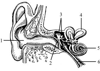
The anatomical elements of the sound-conducting system are the auricle, external auditory canal, tympanic membrane, chain of auditory ossicles, muscles of the tympanic cavity, structures of the vestibule and cochlea (perilymph, endolymph, Reisner's, integumentary and basilar membranes, hairs of sensory cells, secondary tympanic membrane (cochlear window membrane) ) is shown in Fig. 1 general scheme sound transmission systems.
Rice. 1. General diagram of the sound transmission system. The arrows show the direction of the sound wave: 1 - external auditory canal; 2 - supratympanic space; 3 - anvil; 4 - stirrup; 5 — head of the hammer; 6, 10 - scala vestibule; 7, 9 - cochlear duct; 8 - cochlear part of the vestibulocochlear nerve; 11 - scala tympani; 12 - auditory tube; 13 - cochlear window, covered by the secondary tympanic membrane; 14 - window of the vestibule, with the foot plate of the stapes
Each of these elements has specific functions that together provide the primary processing process sound signal- from its “absorption” by the eardrum to its decomposition into frequencies by the structures of the cochlea and its preparation for reception. Removal of any of these elements from the sound transmission process or damage to any of them leads to disruption of the transmission of sound energy, manifested by the phenomenon conductive hearing loss.
Auricle human has retained in a reduced form some useful acoustic functions. Thus, the sound intensity at the level of the external opening of the auditory canal is 3-5 dB higher than in a free sound field. The ears play a certain role in the implementation of the function ototopics And binaural hearing The ears also play a protective role. Due to the special configuration and relief, when air flows over them, diverging vortex flows are formed, preventing air and dust particles from entering the ear canal.
Functional meaning external auditory canal should be considered in two aspects - clinical-physiological and physiological-acoustic. The first is determined by the fact that in the skin of the membranous part of the external auditory canal there are hair follicles, sebaceous and sweat glands, as well as special glands that produce earwax. These formations play a trophic and protective role, preventing the penetration of foreign bodies, insects, and dust particles into the external auditory canal. Earwax, as a rule, is released in small quantities and is a natural lubricant for the walls of the external auditory canal. Being sticky in a “fresh” state, it promotes the adhesion of dust particles to the walls of the membranous-cartilaginous part of the external auditory canal. Drying, it fragments during the act of chewing under the influence of movements in the temporomandibular joint and together with exfoliating particles of the stratum corneum skin and foreign inclusions adhered to it are released out. Earwax has a bactericidal property, as a result of which no microorganisms are found on the skin of the external auditory canal and the eardrum. The length and curvature of the external auditory canal help protect the eardrum from direct injury from a foreign body.
The functional (physiological-acoustic) aspect is characterized by the role played by external auditory canal in conducting sound to the eardrum. This process is not affected by the diameter of the existing or resulting pathological process narrowing of the ear canal, and the length of this narrowing. Thus, with long narrow scar strictures, hearing loss at different frequencies can reach 10-15 dB.
Eardrum is a receiver-resonator of sound vibrations, which, as noted above, has the property of resonating in a wide range of frequencies without significant energy losses. Vibrations of the eardrum are transmitted to the handle of the malleus, then to the incus and stirrup. Vibrations of the foot plate of the stapes are transmitted to the perilymph of the scala vestibularis, which causes vibrations of the main and integumentary membranes of the cochlea. Their vibrations are transmitted to the hair apparatus of auditory receptor cells, in which mechanical energy is transformed into nerve impulses. Vibrations of the perilymph in the scala vestibularis are transmitted through the apex of the cochlea to the perilymph of the scala tympani and then vibrate the secondary tympanic membrane of the cochlear window, the mobility of which ensures the oscillatory process in the cochlea and protects the receptor cells from excessive mechanical stress during loud sounds.
Auditory ossicles combined into a complex lever system that provides increase in strength sound vibrations, necessary to overcome the resting inertia of the perilymph and endolymph of the cochlea and the frictional force of the perilymph in the ducts of the cochlea. The role of the auditory ossicles is also that they, by directly transmitting sound energy to the liquid media of the cochlea, prevent the reflection of the sound wave from the perilymph in the area of the vestibular window.
The mobility of the auditory ossicles is ensured by three joints, two of which ( incus-hammer And anvil-stirrup) are arranged in a typical way. The third joint (the foot plate of the stapes in the window of the vestibule) is only a joint in function; in fact, it is a complex “flap” that performs a dual role: a) ensuring the mobility of the stapes necessary for transmitting sound energy to the structures of the cochlea; b) sealing of the ear labyrinth in the area of the vestibular (oval) window. The element providing these functions is ring connective tissue ligament.
Muscles of the tympanic cavity(the tensor tympani muscle and the stapedius muscle) perform a dual function - protective against strong sounds and adaptive when it is necessary to adapt the sound-conducting system to weak sounds. They are innervated by motor and sympathetic nerves, which in some diseases (myasthenia gravis, multiple sclerosis, various kinds of autonomic disorders) often affects the state of these muscles and may manifest itself in hearing impairment that is not always identifiable.
It is known that the muscles of the tympanic cavity reflexively contract in response to sound stimulation. This reflex comes from receptors in the cochlea. If you apply sound to one ear, a friendly contraction of the muscles of the tympanic cavity occurs in the other ear. This reaction is called acoustic reflex and is used in some hearing research techniques.
There are three types of sound transmission: air, tissue and tube (i.e., through the auditory tube). Air type- this is natural sound conduction, caused by the flow of sound to the hair cells of the spiral organ from the air through the auricle, eardrum and the rest of the sound conduction system. Fabric, or bone, sound conduction is realized as a result of the penetration of sound energy to the moving sound-conducting elements of the cochlea through the tissues of the head. An example of the implementation of bone sound conduction is the tuning fork hearing test technique, in which the handle of a sounding tuning fork is pressed against the mastoid process, crown or other part of the head.
Distinguish compression And inertia mechanism tissue sound conduction. With the compression type, compression and discharge of the liquid media of the cochlea occurs, which causes irritation of the hair cells. With the inertial type, the elements of the sound conducting system, due to the inertial forces developed by their mass, lag behind the rest of the tissues of the skull in their vibrations, resulting in oscillatory movements in the liquid media of the cochlea.
The functions of intracochlear sound conduction include not only the further transmission of sound energy to the hair cells, but also primary spectral analysis sound frequencies, and their distribution among the corresponding sensory elements located on the basilar membrane. With this distribution, a peculiar acoustic-topic principle“cable” transmission of a nerve signal to higher auditory centers, allowing for higher analysis and synthesis of information contained in sound messages.
Auditory reception
Auditory reception is understood as the transformation of the mechanical energy of sound vibrations into electrophysiological nerve impulses, which are a coded expression of an adequate stimulus of the sound analyzer. The receptors of the spiral organ and other elements of the cochlea serve as a generator of biocurrents called cochlear potentials. There are several types of these potentials: resting currents, action currents, microphone potential, summation potential.
Quiescent currents are registered in the absence of a sound signal and are divided into intracellular And endolymphatic potentials. The intracellular potential is recorded in nerve fibers, in hair and supporting cells, in the structures of the basilar and Reissner (reticular) membranes. Endolymphatic potential is recorded in the endolymph of the cochlear duct.
Action currents- These are interfered peaks of bioelectric impulses generated only by the fibers of the auditory nerve in response to sound exposure. The information contained in the action currents is in direct spatial dependence on the location of the neurons stimulated on the main membrane (the theories of hearing by Helmholtz, Bekesy, Davis, etc.). The auditory nerve fibers are grouped into channels, that is, based on their frequency throughput. Each channel is capable of transmitting only a signal of a certain frequency; Thus, if the cochlea is currently affected by low sounds, then only “low-frequency” fibers participate in the process of information transmission, and high-frequency fibers are at rest at this time, i.e., only spontaneous activity is recorded in them. When the cochlea is irritated by a prolonged monophonic sound, the frequency of discharges in individual fibers decreases, which is associated with the phenomenon of adaptation or fatigue.
Snail microphone effect is the result of a response to sound stimulation of only the outer hair cells. Action ototoxic substances And hypoxia lead to suppression or disappearance of the cochlea's microphone effect. However, there is also an anaerobic component in the metabolism of these cells, since the microphonic effect persists for several hours after the death of the animal.
Summation potential owes its origin to the response to sound of the inner hair cells. In the normal homeostatic state of the cochlea, the summation potential recorded in the cochlear duct retains its optimal negative sign, however, slight hypoxia, the action of quinine, streptomycin and a number of other factors that disrupt the homeostasis of the internal media of the cochlea, disrupt the ratio of the magnitudes and signs of the cochlear potentials, at which the summation potential becomes positive.
By the end of the 50s. XX century it was found that in response to sound exposure, certain biopotentials arise in various structures of the cochlea, which give rise to the complex process of sound perception; in this case, action potentials (action currents) arise in the receptor cells of the spiral organ. From a clinical point of view, it seems very important that these cells are highly sensitive to oxygen deficiency, changes in the level of carbon dioxide and sugar in the liquid media of the cochlea, and disturbances in ionic equilibrium. These changes can lead to parabiotic reversible or irreversible pathomorphological changes in the receptor apparatus of the cochlea and to corresponding disorders auditory function.
Otoacoustic emissions. In addition to their main function, the receptor cells of the spiral organ have another amazing property. At rest or under the influence of sound, they come into a state of high-frequency vibration, resulting in the formation of kinetic energy that propagates as a wave process through the tissues of the inner and middle ear and is absorbed by the eardrum. The latter, under the influence of this energy, begins to emit, like a loudspeaker diffuser, a very weak sound in the range of 500-4000 Hz. Otoacoustic emission is not a process of synaptic (nervous) origin, but the result of mechanical vibrations of the hair cells of the spiral organ.
Psychophysiology of hearing
The psychophysiology of hearing considers two main groups of problems: a) measurement threshold of sensation, which is understood as the minimum sensitivity limit sensory system person; b) construction psychophysical scales, reflecting the mathematical dependence or relationship in the “stimulus/response” system for various quantitative values of its components.
There are two forms of sensation threshold − lower absolute threshold of sensation And upper absolute threshold of sensation. By the first we mean the minimum magnitude of the stimulus that causes a response, at which for the first time a conscious sensation of a given modality (quality) of the stimulus arises(in our case - sound). By the second we mean the magnitude of the stimulus at which the sensation of a given modality of the stimulus disappears or changes qualitatively. For example, a powerful sound causes a distorted perception of its tonality or is even extrapolated into the area pain(“pain threshold”).
The magnitude of the sensation threshold depends on the degree of hearing adaptation at which it is measured. When adapting to silence, the threshold decreases; when adapting to a certain noise, it increases.
Subthreshold stimuli those whose magnitude does not cause adequate sensation and does not form sensory perception are called. However, according to some data, subthreshold stimuli, when applied for a sufficiently long time (minutes and hours), can cause “spontaneous reactions” such as causeless memories, impulsive decisions, sudden insights.
Associated with the threshold of sensation are the so-called discrimination thresholds: differential intensity (strength) threshold (DPI or DPS) and differential quality or frequency threshold (DFC). Both of these thresholds are measured as at sequential, and with simultaneous presentation of incentives. When stimuli are presented sequentially, the discrimination threshold can be set if the compared sound intensities and tonality differ by at least 10%. Simultaneous discrimination thresholds, as a rule, are established at the threshold detection of a useful (testing) sound against the background of interference (noise, speech, heteromodal). The method of determining simultaneous discrimination thresholds is used to study the noise immunity of an audio analyzer.
The psychophysics of hearing also considers thresholds of space, locations And time. The interaction of the sensations of space and time gives an integral sense of movement. The sense of movement is based on the interaction of the visual, vestibular and sound analyzers. The location threshold is determined by the spatiotemporal discreteness of the excited receptor elements. Thus, on the basement membrane, a sound of 1000 Hz is displayed approximately in the region of its middle part, and a sound of 1002 Hz is shifted towards the main helix so much that between the sections of these frequencies there is one unexcited cell for which “there was no” corresponding frequency. Therefore, theoretically, the sound location threshold is identical to the frequency discrimination threshold and is 0.2% in the frequency dimension. This mechanism provides an ototopic threshold extrapolated into space in the horizontal plane of 2-3-5°; in the vertical plane this threshold is several times higher.
Psychophysical laws of sound perception form psycho physiological functions sound analyzer. The psychophysiological functions of any sensory organ are understood as the process of the emergence of a sensation specific to a given receptor system when an adequate stimulus acts on it. Psychophysiological methods are based on recording a person’s subjective response to a particular stimulus.
Subjective reactions The hearing organs are divided into two large groups — spontaneous And caused by. The former in their quality are close to the sensations caused by real sound, although they arise “inside” the system, most often due to fatigue of the sound analyzer, intoxication, various local and general diseases. The evoked sensations are caused primarily by the action of an adequate stimulus within given physiological limits. However, they can be provoked by external pathogenic factors (acoustic or mechanical injury ear or auditory centers), then these sensations in their essence approach spontaneous ones.
Sounds are divided into informational And indifferent. Often the latter serve as an obstacle to the former, therefore, in the auditory system there is, on the one hand, a selection mechanism useful information, on the other hand, the interference suppression mechanism. Together they provide one of the most important physiological functions of the sound analyzer - noise immunity.
In clinical studies, only a small part of psychophysiological methods for studying auditory function are used, which are based on only three: a) perception of intensity(strength) of sound, reflected in subjective sensation volume and in the differentiation of sounds by strength; b) frequency perception sound, reflected in the subjective feeling of tone and timbre of sound, as well as in the differentiation of sounds by tonality; V) perception of spatial localization sound source, reflected in the function of spatial hearing (ototopics). All of these functions interact in the natural habitat of humans (and animals), changing and optimizing the process of perception of sound information.
Psychophysiological indicators of hearing function, like any other sense organ, are based on one of essential functions complex biological systems — adaptation.
Adaptation is biological mechanism, with the help of which the body or its individual systems adapt to the energy level of external or internal stimuli acting on them for adequate functioning in the process of their life activity. The process of adaptation of the hearing organ can be implemented in two directions: increased sensitivity to weak sounds or their absence and decreased sensitivity to excessively loud sounds. Increased sensitivity of the hearing organ in silence is called physiological adaptation. The restoration of sensitivity after its decrease, which occurs under the influence of long-acting noise, is called reverse adaptation. The time during which the sensitivity of the hearing organ returns to its original, higher level is called reverse adaptation time(BOA).
The depth of adaptation of the hearing organ to sound exposure depends on the intensity, frequency and duration of the sound, as well as on the time of testing adaptation and the ratio of the frequencies of the influencing and testing sounds. The degree of auditory adaptation is assessed by the magnitude of hearing loss above threshold and by BOA.
Masking is a psychophysiological phenomenon based on the interaction of testing and masking sounds. The essence of masking is that when two sounds of different frequencies are simultaneously perceived, the more intense (louder) sound will mask the weaker one. Two theories compete to explain this phenomenon. One of them gives preference to the neuronal mechanism of the auditory centers, finding confirmation that when exposed to noise in one ear, an increase in the sensitivity threshold in the other ear is observed. Another point of view is based on the peculiarities of biomechanical processes occurring on the basilar membrane, namely during monoaural masking, when the testing and masking sounds are presented in one ear, lower sounds mask higher sounds. This phenomenon is explained by the fact that a “travelling wave” propagating along the basilar membrane from low sounds to the top of the cochlea absorbs similar waves generated from higher frequencies in the lower parts of the basilar membrane, and thus deprives the latter of its ability to resonate at high frequencies. Probably both of these mechanisms take place. The considered physiological functions of the hearing organ underlie all existing methods of its research.
Spatial sound perception
Spatial perception of sound ( ototopics according to V.I. Voyachek) is one of the psychophysiological functions of the hearing organ, thanks to which animals and humans have the ability to determine the direction and spatial position of the sound source. The basis of this function is two-ear (binaural) hearing. Persons with one ear turned off are not able to navigate in space by sound and determine the direction of the sound source. In the clinic, ototopics is important when differential diagnosis peripheral and central lesions of the hearing organ. When the cerebral hemispheres are damaged, various ototopic disorders occur. In the horizontal plane, the ototopic function is performed with greater accuracy than in the vertical plane, which confirms the theory about the leading role of binaural hearing in this function.
Hearing theories
The above psychophysiological properties of the sound analyzer are, to one degree or another, explained by a number of hearing theories developed in late XIX- early 20th century
Helmholtz's resonance theory explains the emergence of tonal hearing by the phenomenon of resonating the so-called strings of the main membrane at different frequencies: short fibers of the main membrane located in the lower helix of the cochlea resonate to high sounds, fibers located in the middle helix of the cochlea resonate to medium frequencies, and to low frequencies in the upper helix , where the longest and most relaxed fibers are located.
Bekesy traveling wave theory is based on hydrostatic processes in the cochlea, which, with each oscillation of the foot plate of the stapes, causes deformation of the main membrane in the form of a wave running towards the apex of the cochlea. At low frequencies The traveling wave reaches a section of the main membrane located at the apex of the cochlea, where the long “strings” are located; at high frequencies, the waves cause bending of the main membrane in the main helix, where the short “strings” are located.
Theory of P. P. Lazarev explains spatial perception separate frequencies along the main membrane by the unequal sensitivity of the hair cells of the spiral organ to different frequencies. This theory was confirmed in the works of K. S. Ravdonik and D. I. Nasonov, according to which living cells of the body, regardless of their affiliation, react with biochemical changes to sound irradiation.
Theories about the role of the main membrane in the spatial discrimination of sound frequencies were confirmed in studies with conditioned reflexes in the laboratory of I. P. Pavlov. In these studies, a conditioned food reflex was developed to different frequencies, which disappeared after the destruction of different parts of the main membrane responsible for the perception of certain sounds. V.F. Undritz studied the biocurrents of the snail, which disappeared when various sections of the main membrane were destroyed.
Otorhinolaryngology. IN AND. Babiyak, M.I. Govorun, Ya.A. Nakatis, A.N. Pashchinin
Consists of the outer, middle and inner ear. Average and inner ear are located inside the temporal bone.
Outer ear consists of the auricle (collects sounds) and the external auditory canal, which ends in the eardrum.
Middle ear- This is a chamber filled with air. It contains the auditory ossicles (hammer, incus and stapes), which transmit vibrations from the eardrum to the membrane of the oval window - they amplify the vibrations 50 times. The middle ear is connected to the nasopharynx via the Eustachian tube, through which the pressure in the middle ear is equalized with atmospheric pressure.
In the inner ear there is a cochlea - a fluid-filled bone canal twisted in 2.5 turns, blocked by a longitudinal septum. On the septum there is an organ of Corti containing hair cells - these are auditory receptors that convert sound vibrations into nerve impulses.
Ear work: When the stapes presses on the membrane of the oval window, the column of fluid in the cochlea moves, and the membrane of the round window protrudes into the middle ear. The movement of the fluid causes the hairs to touch the integumentary plate, causing the hair cells to become excited.
Vestibular apparatus: In the inner ear, in addition to the cochlea, there are semicircular canals and vestibular sacs. Hair cells in the semicircular canals sense fluid movement and respond to acceleration; hair cells in the sacs sense the movement of the otolith pebble attached to them and determine the position of the head in space.
Establish a correspondence between the structures of the ear and the sections in which they are located: 1) outer ear, 2) middle ear, 3) inner ear. Write the numbers 1, 2 and 3 in the correct order.
A) auricle
B) oval window
B) snail
D) stirrup
D) Eustachian tube
E) hammer
Answer
Establish a correspondence between the function of the hearing organ and the section that performs this function: 1) middle ear, 2) inner ear
A) conversion of sound vibrations into electrical ones
B) amplification of sound waves due to vibrations of the auditory ossicles
B) equalization of pressure on the eardrum
D) conducting sound vibrations due to the movement of liquid
D) irritation of auditory receptors
Answer
1. Establish the sequence of sound wave transmission to the auditory receptors. Write down the corresponding sequence of numbers.
1) vibrations of the auditory ossicles
2) vibrations of fluid in the cochlea
3) vibrations of the eardrum
4) irritation of auditory receptors
Answer
2. Establish the correct sequence of passage of a sound wave in the human hearing organ. Write down the corresponding sequence of numbers.
1) eardrum
2) oval window
3) stirrup
4) anvil
5) hammer
6) hair cells
Answer
3. Establish the sequence in which sound vibrations are transmitted to the receptors of the hearing organ. Write down the corresponding sequence of numbers.
1) Outer ear
2) Membrane of the oval window
3) Auditory ossicles
4) Eardrum
5) Fluid in the cochlea
6) Hearing receptors
Answer
4. Establish the sequence of arrangement of the structures of the human ear, starting with the one that captures the sound wave. Write down the corresponding sequence of numbers.
1) oval window of the cochlea of the inner ear
2) external auditory canal
3) eardrum
4) auricle
5) auditory ossicles
6) organ of Corti
Answer
5. Establish the sequence of transmission of sound vibrations to the receptors of the human hearing organ. Write down the corresponding sequence of numbers.
1) external auditory canal
2) oval window membrane
3) auditory ossicles
4) eardrum
5) fluid in the cochlea
6) hair cells of the cochlea
Answer
1. Select three correctly labeled captions for the drawing “Structure of the Ear.”
1) external auditory canal
2) eardrum
3) auditory nerve
4) stirrup
5) semicircular canal
6) snail
Answer
2. Select three correctly labeled captions for the drawing “Structure of the Ear.” Write down the numbers under which they are indicated.
1) ear canal
2) eardrum
3) auditory ossicles
4) auditory tube
5) semicircular canals
6) auditory nerve
Answer
4. Select three correctly labeled captions for the drawing “Structure of the Ear.”
1) auditory ossicles
2) facial nerve
3) eardrum
4) auricle
5) middle ear
6) vestibular apparatus
Answer
1. Set the sequence of sound transmission in the hearing analyzer. Write down the corresponding sequence of numbers.
1) vibration of the auditory ossicles
2) fluid vibrations in the cochlea
3) generation of a nerve impulse
5) transmission of nerve impulses auditory nerve in the temporal lobe of the cortex cerebral hemispheres
6) vibration of the oval window membrane
7) vibration of hair cells
Answer
2. Establish the sequence of processes occurring in the auditory analyzer. Write down the corresponding sequence of numbers.
1) transmission of vibrations to the membrane of the oval window
2) capturing the sound wave
3) irritation of receptor cells with hairs
4) vibration of the eardrum
5) movement of fluid in the cochlea
6) vibration of the auditory ossicles
7) the occurrence of a nerve impulse and its transmission along the auditory nerve to the brain
Answer
3. Establish the sequence of processes of passage of a sound wave in the organ of hearing and a nerve impulse in the auditory analyzer. Write down the corresponding sequence of numbers.
1) movement of fluid in the cochlea
2) transmission of sound waves through the malleus, incus and stapes
3) transmission of nerve impulses along the auditory nerve
4) vibration of the eardrum
5) conduction of sound waves through the external auditory canal
Answer
4. Establish the path of the sound wave of a car siren that a person will hear, and the nerve impulse that occurs when it sounds. Write down the corresponding sequence of numbers.
1) snail receptors
2) auditory nerve
3) auditory ossicles
4) eardrum
5) auditory cortex
Answer
Choose the one that suits you best correct option. The auditory analyzer receptors are located
1) in the inner ear
2) in the middle ear
3) on the eardrum
4) in the auricle
Answer
Choose one, the most correct option. The sound signal is converted into nerve impulses in
1) snail
2) semicircular canals
3) eardrum
4) auditory ossicles
Answer
Choose one, the most correct option. In the human body, an infection from the nasopharynx enters the middle ear cavity through
1) oval window
2) larynx
3) auditory tube
4) inner ear
Answer
Establish a correspondence between the parts of the human ear and their structure: 1) outer ear, 2) middle ear, 3) inner ear. Write the numbers 1, 2, 3 in the order corresponding to the letters.
A) includes auricle and external auditory canal
B) includes the cochlea, which contains the initial section of the sound-receiving apparatus
B) includes three auditory ossicles
D) includes the vestibule with three semicircular canals, which contain the balance apparatus
D) a cavity filled with air communicates through the auditory tube with the pharyngeal cavity
E) the inner end is covered by the eardrum
Answer
Establish a correspondence between the characteristics and analyzers of a person: 1) visual, 2) auditory. Write numbers 1 and 2 in the order corresponding to the letters.
A) perceives mechanical vibrations environment
B) includes rods and cones
IN) central department is located in temporal lobe cerebral cortex
D) the central section is located in the occipital lobe of the cerebral cortex
D) includes the organ of Corti
Answer
Select three correctly labeled captions for the figure “Structure of the vestibular apparatus.” Write down the numbers under which they are indicated.
1) Eustachian tube
2) snail
3) calcareous crystals
4) hair cells
5) nerve fibers
6) inner ear
Answer
Choose one, the most correct option. Pressure on the eardrum equal to atmospheric pressure from the middle ear is provided in humans
1) auditory tube
2) auricle
3) membrane of the oval window
4) auditory ossicles
Answer
Choose one, the most correct option. Receptors that determine the position of the human body in space are located in
1) membrane of the oval window
2) eustachian tube
3) semicircular canals
4) middle ear
Answer
Choose three correct answers out of six and write down the numbers under which they are indicated. The hearing analyzer includes:
1) auditory ossicles
2) receptor cells
3) auditory tube
4) auditory nerve
5) semicircular canals
6) temporal lobe cortex
Answer
Choose three correct answers out of six and write down the numbers under which they are indicated. What is included in the auditory sensory system?
1) semicircular canals
2) bone labyrinth
3) snail receptors
4) auditory tube
5) vestibulocochlear nerve
6) temporal zone of the cerebral cortex
Answer
Choose three correct answers out of six and write down the numbers under which they are indicated. The middle ear in the human hearing organ includes
1) receptor apparatus
2) anvil
3) auditory tube
4) semicircular canals
5) hammer
6) auricle
Answer
Choose three correct answers out of six and write down the numbers under which they are indicated. What should be considered true signs of the human hearing organ?
1) The external auditory canal is connected to the nasopharynx.
2) Sensitive hair cells are located on the membrane of the cochlea of the inner ear.
3) The middle ear cavity is filled with air.
4) The middle ear is located in the labyrinth of the frontal bone.
5) The outer ear detects sound vibrations.
6) The membranous labyrinth amplifies sound vibrations.
Answer
Establish a correspondence between the characteristics and sections of the hearing organ presented in the diagram. Write numbers 1 and 2 in the order corresponding to the letters.
A) amplifies sound vibrations
B) converts mechanical vibrations into nerve impulses
B) contains auditory ossicles
D) filled with incompressible fluid
D) contains the organ of Corti
E) participates in equalizing air pressure
Answer
© D.V. Pozdnyakov, 2009-2019
Human organism. The structure and vital functions of organs and organ systems. Human hygiene.
Task 14: human body. The structure and vital functions of organs and organ systems. Human hygiene.
(sequencing)
1. Establish the correct sequence of passage of a sound wave and a nerve impulse through the auditory analyzer from a shot to the cerebral cortex. Write down the corresponding sequence of numbers in the table.
- Shot sound
- Auditory cortex
- Auditory ossicles
- Snail receptors
- Auditory nerve
- Eardrum
Answer: 163452.
2. Establish the sequence of bends of the human spine, starting with the head. Write down the corresponding sequence of numbers in the table.
- Lumbar
- Cervical
- Sacral
- Chest
Answer: 2413.
3. Establish the correct sequence of actions to stop arterial bleeding from radial artery. Write down the corresponding sequence of numbers in the table.
- Deliver the victim to a medical facility
- Free your forearm from clothing
- Place above the wound site soft cloth, and put a rubber band on top
- Tie the tourniquet in a knot or tie it with a wooden stick-twist
- Attach a piece of paper to the tourniquet indicating the time of its application
- Place a sterile gauze bandage on the wound surface and bandage it
Answer: 234651.
4. Establish the correct sequence of movement of arterial blood in a person, starting from the moment it is saturated with oxygen in the capillaries of the pulmonary circle. Write down the corresponding sequence of numbers in the table.
- Left ventricle
- Left atrium
- Veins of the small circle
- Arteries great circle
- Small circle capillaries
Answer: 53214.
5. Establish the correct sequence of elements of the reflex arc of the cough reflex in a person. Write down the corresponding sequence of numbers in the table.
- Executive neuron
- Laryngeal receptors
- Center of the medulla oblongata
- Sensory neuron
- Contraction of respiratory muscles
Answer: 24315.
6. Establish the correct sequence of processes occurring during blood clotting in humans. Write down the corresponding sequence of numbers in the table.
- Prothrombin formation
- Blood clot formation
- Fibrin formation
- Damage to the vessel wall
- Effect of thrombin on fibrinogen
Answer: 41532.
7. Establish the correct sequence of digestive processes in humans. Write down the corresponding sequence of numbers in the table.
- Supply of nutrients to organs and tissues of the body
- Transfer of food to the stomach and its digestion gastric juice
- Grinding food with teeth and changing it under the influence of saliva
- Absorption of amino acids into the blood
- Digestion of food in the intestines under the influence of intestinal juice, pancreatic juice and bile
Answer: 32541.
8. Establish the correct sequence of elements of the reflex arc of the human knee reflex. Write down the corresponding sequence of numbers in the table.
- Sensory neuron
- Motor neuron
- Spinal cord
- Quadriceps femoris
- Tendon receptors
Answer: 51324.
9. Set the correct sequence of dice upper limb starting from the shoulder girdle. Write down the corresponding sequence of numbers in the table.
- Carpal bones
- Metacarpal bones
- Phalanges of fingers
- Radius
- Brachial bone
Answer: 54123.
10. Establish the correct sequence of digestive processes in humans. Write down the corresponding sequence of numbers in the table.
- Breakdown of polymers into monomers
- Swelling and partial breakdown of proteins
- Absorption of amino acids and glucose into the blood
- Beginning of starch breakdown
- Intensive water absorption
Answer: 42135.
11. Establish the sequence of stages of inflammation when microbes penetrate (for example, when damaged by a splinter). Write down the corresponding sequence of numbers in the table.
- Destruction of pathogens
- Redness of the affected area: capillaries dilate, blood flows in, local temperature rises, sensation of pain
- Leukocytes arrive with blood to the inflamed area
- A powerful protective layer of leukocytes and macrophages is formed around the accumulation of microbes
- Concentration of microbes in the affected area
Answer: 52341.
12. Establish the sequence of stages of the human cardiac cycle after a pause (that is, after the chambers are filled with blood). Write down the corresponding sequence of numbers in the table.
- Blood supply to the superior and inferior vena cava
- The blood gives off nutrients and oxygen and receives metabolic products and carbon dioxide
- Blood flow into arteries and capillaries
- Contraction of the left ventricle, blood flow into the aorta
- Blood flow into the right atrium of the heart
Answer: 43215.
13. Establish the sequence of location of the human airways. Write down the corresponding sequence of numbers in the table.
- Bronchi
- Nasopharynx
- Larynx
- Trachea
- Nasal cavity
Answer: 52341.
14. Arrange in the correct order the sequence of the bones of the leg skeleton from top to bottom. Write down the corresponding sequence of numbers in the table.
- Metatarsus
- Femur
- Shin
- Tarsus
- Phalanges of fingers
Answer: 23415.
15. Signs of fatigue during static work are recorded in the experiment of holding a load in an arm extended strictly horizontally to the side. Establish the sequence of manifestation of signs of fatigue in this experiment. Write down the corresponding sequence of numbers in the table.
- Hand tremors, loss of coordination, staggering, facial flushing, sweating
- The hand with the load goes down
- The hand drops, then jerks back to its original place.
- Recovery
- The hand with the load is motionless
Answer: 53124.
16. Establish the sequence of stages of carbon dioxide transport from brain cells to the lungs. Write down the corresponding sequence of numbers in the table.
- Pulmonary arteries
- Right atrium
- Jugular vein
- Pulmonary capillaries
- Right ventricle
- Superior vena cava
- Brain cells
Answer: 7362514.
17. Establish the sequence of processes in the cardiac cycle. Write down the corresponding sequence of numbers in the table.
- Flow of blood from the atria into the ventricles
- Diastole
- Atrial contraction
- Closing of leaflet valves and opening of semilunar valves
- Blood supply to the aorta and pulmonary arteries
- Ventricular contraction
- Blood from the veins enters the atria and partially flows into the ventricles
Answer: 3164527.
18. Establish the sequence of processes occurring during the regulation of the work of internal organs. Write down the corresponding sequence of numbers in the table.
- The hypothalamus receives a signal from the internal organ
- Endocrine gland produces a hormone
- The pituitary gland produces tropic hormones
- The functioning of the internal organ changes
- Transport of tropic hormones to the endocrine glands
- Release of neurohormones
Answer: 163524.
19. Establish the sequence of location of the intestinal sections in humans. Write down the corresponding sequence of numbers in the table.
- Skinny
- Sigmoid
- Blind
- Straight
- Colon
- duodenum
- Ileum
Answer: 6173524.
20. Establish the sequence of processes occurring in the human female reproductive system in the event of pregnancy. Write down the corresponding sequence of numbers in the table.
- Attachment of the embryo to the wall of the uterus
- Release of the egg into the fallopian tube - ovulation
- Maturation of the egg in the graph vesicle
- Multiple divisions of the zygote, formation of the germinal vesicle - blastula
- Fertilization
- Movement of the egg due to the movement of cilia ciliated epithelium fallopian tube
- Placentation
Answer: 3265417.
21. Establish the sequence of periods of development in a person after birth. Write down the corresponding sequence of numbers in the table.
- Newborn
- Puberty
- Early childhood
- Teenage
- Preschool
- Chest
- Youthful
Answer: 1635247.
22. Establish the sequence of information transfer along the links of the reflex arc of the ciliary reflex. Write down the corresponding sequence of numbers in the table.
- Transfer of excitation to the orbicularis oculi muscle, which closes the eyelids
- Transmission of a nerve impulse along the axon of a sensory neuron
- Transmission of information to the executive neuron
- Receiving information by an interneuron and transmitting it to the medulla oblongata
- The emergence of excitation in the center of the blink reflex
- Getting a speck in the eye
Answer: 624531.
23. Establish the sequence of sound wave propagation in the organ of hearing. Write down the corresponding sequence of numbers in the table.
- Hammer
- Oval window
- Eardrum
- Stapes
- Fluid in the cochlea
- Anvil
Answer: 316425.
24. Establish the sequence of movement of carbon dioxide in humans, starting from the cells of the body. Write down the corresponding sequence of numbers in the table.
- Superior and inferior vena cava
- Body cells
- Right ventricle
- Pulmonary arteries
- Right atrium
- Capillaries of the systemic circulation
- Alveoli
Answer: 2615437.
25. Set the sequence of information transfer in olfactory analyzer. Write down the corresponding sequence of numbers in the table.
- Irritation of the cilia of olfactory cells
- Analysis of information in the olfactory zone of the cerebral cortex
- Transmission of olfactory impulses to the subcortical nuclei
- When inhaled, odorous substances enter the nasal cavity and dissolve in mucus.
- The emergence of olfactory sensations, which also have emotional coloring
- Transmission of information along the olfactory nerve
Answer: 416235.
26. Set the sequence of steps fat metabolism in humans. Write down the corresponding sequence of numbers in the table.
- Emulsification of fats under the influence of bile
- Uptake of glycerol and fatty acids by intestinal villus epithelial cells
- Entry of human fat into the lymphatic capillary and then into the fat depot
- Fat intake from food
- Synthesis of human fat in epithelial cells
- Breakdown of fats into glycerol and fatty acids
Answer: 416253.
27. Establish the sequence of steps for preparing tetanus serum. Write down the corresponding sequence of numbers in the table.
- Administration of tetanus toxoid to the horse
- Developing lasting immunity in horses
- Preparation of antitetanus serum from purified blood
- Purifying the horse's blood - removing blood cells, fibrinogen and proteins from it
- Repeated administration of tetanus toxoid to a horse at regular intervals with increasing doses
- Taking blood from a horse
Answer: 152643.
28. Establish the sequence of processes occurring during the development of a conditioned reflex. Write down the corresponding sequence of numbers in the table.
- Presentation of a conditional signal
- Multiple repetition
- Development of a conditioned reflex
- The emergence of a temporary connection between two foci of excitation
- Unconditional reinforcement
- The emergence of foci of excitation in the cerebral cortex
Answer: 156243.
29. Establish the sequence of passage through the organs of the human respiratory system of a labeled oxygen molecule that penetrates into the lungs during inhalation. Write down the corresponding sequence of numbers in the table.
- Nasopharynx
- Bronchi
- Larynx
- Nasal cavity
- Lungs
- Trachea
Answer: 413625.
30. Establish the path that nicotine travels through the blood from the pulmonary alveoli to the brain cells. Write down the corresponding sequence of numbers in the table.
- Left atrium
- Carotid artery
- Pulmonary capillary
- Brain cells
- Aorta
- Pulmonary veins
- Left ventricle
Answer: 3617524.
Biology. Preparation for the Unified State Exam 2018. 30 training options based on the 2018 demo version: educational and methodological manual/A. A. Kirilenko, S. I. Kolesnikov, E. V. Dadenko; edited by A. A. Kirilenko. - Rostov n/d: Legion, 2017. - 624 p. - (Unified State Examination).
1. Establish the correct sequence of nerve impulse transmission along the reflex arc. Write down the corresponding sequence of numbers in the table.
- Interneuron
- Receptor
- Effector neuron
- Sensory neuron
- Working body
Answer: 24135.
2. Establish the correct sequence of passage of a portion of blood from the right ventricle to the right atrium. Write down the corresponding sequence of numbers in the table.
- Pulmonary vein
- Left ventricle
- Pulmonary artery
- Right ventricle
- Right atrium
- Aorta
Answer: 431265.
3. Establish the correct sequence of breathing processes in a person, starting with an increase in the concentration of CO2 in the blood. Write down the corresponding sequence of numbers in the table.
- Increasing oxygen concentration
- Increased CO2 concentration
- Excitation of chemoreceptors of the medulla oblongata
- Exhalation
- Contraction of the respiratory muscles
Answer: 346125.
4. Establish the correct sequence of processes occurring during blood clotting in humans. Write down the corresponding sequence of numbers in the table.
- Blood clot formation
- Interaction of thrombin with fibrinogen
- Platelet destruction
- Damage to the vessel wall
- Fibrin formation
- Activation of prothrombin
Answer: 436251.
5. Establish the correct sequence of first aid measures medical care with bleeding from the brachial artery. Write down the corresponding sequence of numbers in the table.
- Apply a tourniquet to the tissue above the wound site
- Take the victim to the hospital
- Place a note under the tourniquet indicating the time it was applied.
- Press the artery to the bone with your finger
- Apply a sterile dressing over the tourniquet
- Check that the tourniquet is applied correctly by feeling the pulse
Answer: 416352.
6. Establish the correct sequence of first aid measures for a drowning person. Write down the corresponding sequence of numbers in the table.
- Press rhythmically on the back to remove water from respiratory tract
- Deliver the victim to medical institution
- Place the victim face down on the thigh of the rescuer's bent leg
- Perform mouth-to-mouth artificial respiration while holding your nose
- Clean the victim’s nasal and oral cavities from dirt and mud
Answer: 53142.
7. Establish the sequence of processes that occur during inhalation. Write down the corresponding sequence of numbers in the table.
- The lungs, following the walls of the chest cavity, expand
- The appearance of a nerve impulse in respiratory center
- Air flows through the airways into the lungs - inhalation occurs
- When the external intercostal muscles contract, the ribs rise
- The volume of the chest cavity increases
Answer: 24513.
8. Establish the sequence of processes of passage of a sound wave in the organ of hearing and a nerve impulse in the auditory analyzer. Write down the corresponding sequence of numbers in the table.
- Movement of fluid in the cochlea
- Transmission of sound waves through the malleus, incus and stapes
- Transmission of nerve impulses along the auditory nerve
- Eardrum vibration
- Conduction of sound waves through the external auditory canal
Answer: 54213.
9. Establish the sequence of stages of formation and movement of urine in the human body. Write down the corresponding sequence of numbers in the table.
- Accumulation of urine in the renal pelvis
- Reabsorption from nephron tubules
- Blood plasma filtration
- The flow of urine through the ureter into the bladder
- Movement of urine through the collecting ducts of the pyramids
Answer: 32514.
10. Establish the sequence of processes occurring in digestive system humans when digesting food. Write down the corresponding sequence of numbers in the table.
- Grinding, mixing food and primary breakdown of carbohydrates
- Absorption of water and breakdown of fiber
- Protein breakdown in an acidic environment under the influence of pepsin
- Absorption of amino acids and glucose into the blood through the villi
- Passing a food bolus through the esophagus
Answer: 15342.
11. Establish the sequence of processes occurring in the human digestive system. Write down the corresponding sequence of numbers in the table.
- Protein breakdown by pepsin
- Breakdown of starch in an alkaline environment
- Digestion of fiber by symbiotic bacteria
- Movement food bolus along the esophagus
- Absorption of amino acids and glucose through the villi
Answer: 24153.
12. Establish the sequence of thermoregulation processes in humans during muscular work. Write down the corresponding sequence of numbers in the table.
- Signal transmission along the motor pathway
- Relaxation of blood vessel muscles
- Impact low temperatures to skin receptors
- Increased heat transfer from the surface of blood vessels
Rice. 5.18. Sound wave.
p - sound pressure; t - time; l is the wavelength.
hearing is sound, therefore, to highlight the main functional features of the system, it is necessary to become familiar with some concepts of acoustics.
Basic physical concepts of acoustics. Sound is mechanical vibrations of an elastic medium, propagating in the form of waves in air, liquid and solids. The source of sound can be any process that causes a local change in pressure or mechanical stress in the medium. From a physiological point of view, sound is understood as mechanical vibrations that, acting on auditory receptor, cause a certain physiological process in it, perceived as a sensation of sound.
The sound wave is characterized by sinusoidal, i.e. periodic, oscillations (Fig. 5.18). When propagating in a certain medium, sound is a wave with phases of condensation (densification) and rarefaction. There are transverse waves - in solids, and longitudinal waves - in air and liquid media. The speed of propagation of sound vibrations in air is 332 m/s, in water - 1450 m/s. Identical states of a sound wave - areas of condensation or rarefaction - are called phases. The distance between the middle and extreme positions of the oscillating body is called amplitude of oscillations, and between identical phases - wavelength. The number of oscillations (compression or rarefaction) per unit time is determined by the concept sound frequencies. The unit of sound frequency is hertz(Hz), indicating the number of vibrations per second. Distinguish high frequency(high) and low frequency(low) sounds. Low sounds, in which the phases are far apart from each other, have a long wavelength, high sounds with close phases have a small (short) wavelength.
Phase And wavelength are important in the physiology of hearing. Thus, one of the conditions for optimal hearing is the arrival of a sound wave at the windows of the vestibule and cochlea in different phases and this is anatomically ensured sound conducting system middle ear. High-pitched sounds with a short wavelength vibrate a small (short) column of labyrinthine fluid (perilymph) at the base of the cochlea (here they
are perceived), low ones - with a long wavelength - extend to the apex of the cochlea (here they are perceived). This circumstance is important for understanding modern theories of hearing.
Based on the nature of the oscillatory movements, they are distinguished:
Pure tones;
Complex tones;
Harmonic (rhythmic) sine waves create a clear, simple sound tone. An example would be the sound of a tuning fork. Non-harmonic sound, different from simple sounds complex structure, is called noise. The frequencies of the various oscillations that create the noise spectrum are related to the frequency of the fundamental tone chaotically, like various fractional numbers. The perception of noise is often accompanied by unpleasant subjective sensations.
The ability of a sound wave to bend around obstacles is called diffraction. Low sounds with a long wavelength have better diffraction than high sounds with a short wavelength. The reflection of a sound wave from obstacles encountered in its path is called echo. The repeated reflection of sound in enclosed spaces from various objects is called reverberation. The phenomenon of superposition of a reflected sound wave on a primary sound wave is called "interference". In this case, an increase or decrease in sound waves may be observed. When sound passes through the external auditory canal, interference occurs and the sound wave is amplified.
The phenomenon when a sound wave of one vibrating object causes vibrational movements of another object is called resonance. Resonance can be sharp when the natural period of oscillation of the resonator coincides with the period of the acting force, and blunt if the periods of oscillation do not coincide. With an acute resonance, the oscillations decay slowly, with a dull resonance, they decay quickly. It is important that vibrations of the ear structures that conduct sounds decay quickly; this eliminates the distortion of external sound, so a person can quickly and consistently receive more and more new sound signals. Some structures of the cochlea have a sharp resonance, and this helps to distinguish between two closely spaced frequencies.
Basic properties of the auditory analyzer. These include the ability to distinguish between pitch, volume and timbre. The human ear perceives sound frequencies from 16 to 20,000 Hz, which is 10.5 octaves. Oscillations with a frequency of less than 16 Hz are called infrasound, and above 20,000 Hz - Ultrasound. Infrasound and ultrasound under normal conditions