
Symptoms and methods of treatment of Shinz disease in children. Osteochondropathy of the calcaneus in children treatment
- Appearances and symptoms of osteochondropathy
- Characteristic signs and development of the disease
- It is necessary to get rid of the disease in a complex
- Getting rid of osteochondropathy folk methods and prevention
Osteochondropathy of the calcaneus is a disease characteristic of the female half of humanity. In particular, adolescent girls suffer from it. Affects one or both legs at once.
Haglund's disease, or Haglund's deformity - as it is also called in the medical world, since the first description of the disease was given in 1907 by the Swedish orthopedic surgeon P. Haglund. It is believed that in young children, osteochondropathy occurs infrequently, but can also occur at the age of 9 years. It is possible to cure the disease completely without resorting to surgical methods . With only one caveat that getting rid of the disease will begin in the early stages.
Appearances and symptoms of osteochondropathy
Strong physical exertion or injury can lead to an illness. Damage interferes with normal blood circulation, while bone and cartilage begin to break down. It brings pain, there is swelling, sometimes quite strong. The heel pulsates, becomes hot. Local temperature sometimes lasts long enough and affects the calcaneal tendon. Osteochondropathy can go away without treatment, but in the acute period, pain relief measures must be taken, and doctors recommend carefully monitoring the patient's condition, even if the symptoms of the disease have passed.
In addition to the calcaneus, the lesion can affect the thigh, spine, and tibia. Scientists cannot say exactly why this disease occurs. It is known that mobile people experience this condition more often than others. There are a number of other factors that are considered harbingers of osteochondropathy: 
- congenital pathology.
- Heredity.
- Hormonal deviations.
- Disturbances in body systems, in particular fast growth bone or muscle tissue.
- Poor absorption of calcium.
- Injuries.
- Excessive exertion, especially as a result of long running and jumping.
Loads lead to compression of small vessels of spongy bones. This impairs local blood flow, there is insufficient tissue replenishment. Osteochondropathy of the calcaneal tuber develops in 5 stages:
- aseptic necrosis, which is the necrosis of bone tissue cells;
- fragmentation and fracture;
- tissue resorption;
- recovery process;
- osteoarthritis, inflammation if left untreated.
The tubercle of the calcaneus does not allow a person to move normally, while I want to put the foot on the toe. The disease can be diagnosed by X-ray: the image shows seals and uncharacteristic roughness on the tubercle of the heel bone. Often the symptoms of the disease completely coincide with those characteristic of osteochondropathy, but the picture clearly shows that the patient has. Therefore, it is very important not to self-medicate at the first manifestations of pain in the foot and heel - only an orthopedist after the examination is able to prescribe the necessary course of therapy.
Back to index
Characteristic signs and development of the disease
 Pain in the heel does not appear when the leg is at rest, but when moving or if you press your fingers on the area of \u200b\u200bthe foot that bothers. In other diseases, such as bursitis, tumor, bone tuberculosis, periostitis, completely different symptoms are observed. The pain lasts constantly, the heel suffering from osteochondropathy does not turn red, but only swells.
Not less than important feature- the image shows tissue in those parts of the bone that are affected by the disease. They are displaced to the side, the contour of the bone is uneven and strongly pronounced, quite different from the healthy state.
The disease develops rather slowly. The manifestations of pathology and the rate of the course of the disease depend on the age of the patient and his physical condition. Sometimes it takes several months or even years to reach the main stage. It all starts with reduced muscle tone, rapid fatigue of the patient. The condition improves after sleep, but returns with intense exercise. Celebrated and hypersensitivity skin in the area of the calcaneal tubercle.
Contact your doctor as soon as possible. On the initial stage even simple anesthesia can significantly improve the picture.
Back to index
It is necessary to get rid of the disease in a complex
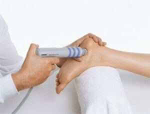
Conservative treatment is not always effective, orthopedists are sure. But you need to start with him. It is important not to put a load on the sore heel (she needs rest), to observe bed rest, but to move around with the help of crutches. The affected areas are chipped with novocaine. The doctor prescribes physiotherapy:
- electrophoresis of novocaine together with analgin;
- microwave therapy;
- applications with ozocerite;
- specialized baths and compresses.
 Painkillers are also shown in the form of tablets. It is important to drink vitamin complex with the content of vitamins of group B. Vitamins B6 and B12 are especially important.
With a strong pain syndrome and if the main treatment does not work, orthopedists recommend cutting the nerve in the diseased area. In particular, the nerves leading to the heel and the tibial nerve are isolated. Neurotomy will allow the patient to lead a full-fledged lifestyle, move normally and even give small physical exertion to the feet with sports training. But such an operation leads to a loss not only of sensitivity in the heels, but the skin in this place ceases to feel any touch.
If all the manipulations were carried out in a timely manner, the calcaneus will recover and the pain will not return. But when self-treatment takes place, the tubercle of the bone will stick out. This will be reflected in inconvenience when wearing shoes of any type (except slippers without backs). Surgical intervention and specialized orthopedic shoes will come to the rescue. Subsequently, it is necessary to continue the use of arch support insoles. A medium-intensity foot massage and a general massage are shown.
Osteochondropathy of the calcaneus in children is a dangerous degenerative disease that leads to severe degenerative damage to bone tissue. As a result of the disease, the bone almost completely loses its strength and, as a result, the ability to perform its musculoskeletal functions. Depending on the places of localization, the degenerative process, its stage, the danger and methods of treatment change.
Causes of pathology
Osteochondropathy of the calcaneus remains a mystery to specialists to this day, because the mechanism provoking its development has not yet been identified. It is believed that an important role in this process is played by such pathogenic factors as:
- excessive stress on the tendons of the feet;
- microtrauma;
- systemic failures in the body.
But, since the body of babies is often, due to their age, protected from most known pathogenic factors, the mechanism of the development of the disease is not fully understood.
degenerative change bone structure legs in a child is often called the result of traumatic loads. Many medical professionals agree that injuries in the area knee joint or heel bone can cause the development of osteochondropathy. Damaged tissue regenerates over time. It is able to activate a similar degenerative process in the "intact" bone adjacent to the diseased area. It is not uncommon for the disease to:
- be inherited if the disease is regularly detected in close relatives;
- be the result of misconduct endocrine system and poor absorption of calcium;
- indicate the presence of active pathological processes in the body;
- be the result of a circulatory disorder - the most commonly cited cause.
Depending on the age, the pathology has its own localization, in which it is usually diagnosed for the first time.
Adult patients and adolescents most often suffer from damage to the hyaline cartilage, older children - to the epiphyseal zone. And in babies, the central part of the bone usually undergoes a degenerative process. But regardless of its location, without proper care, the disease causes enormous, sometimes irreversible harm to their body.
Stages of development

Osteochondropathy of the calcaneal tuber, regardless of the age of the patient, can be caused by circulatory disorders. But, whether this is true or not, further forecasts in the absence of a proper approach are disappointing. The disease develops gradually and consists of several stages:
- aseptic necrosis of bone tissue;
- fracture and partial fragmentation;
- resorption of necrotic bone tissue;
- restoration of damaged areas - with the correct diagnosis;
- inflammation or the development of deforming osteoarthritis - in the absence of treatment.
Aseptic necrotization of the calcaneal apophysis is the first stage of the disease and includes several processes.
Being the result of a violation of the normal nutrition of tissues due to damage to blood vessels or for other reasons, it is a kind of tissue death of the heel bones from starvation. This process is quite extended in time, and almost never gives the patient discomfort. The duration of this stage can often be at least six months.
Six months the development of osteochondropathy does not bring the baby any serious problems or discomfort. Given that to identify the disease on initial stage very difficult, parents may not be aware that a pathological process has begun in the child's body. Its presence can be found in the second stage of the disease. It also lasts an average of about six months. But unlike the first, it allows, with the help of an X-ray examination, to see violations in the bone structure that have become the result of a degenerative process.
The third stage is characterized by fragmentation of the heel bones. In the bone tissue destroyed by necrosis develops inflammatory process, damaged areas are gradually dissolved by the body. The uninjured are tied together by cords. connective tissue. X-ray allows you to see that the calcaneus has turned into a structure consisting of separate parts, interconnected by "threads" of connective tissue. It is very important at this stage to ensure the correct treatment, since the further health of the baby's legs depends on it. One of the prerequisites is often the restriction of their mobility in order for the regeneration process to proceed more efficiently.
Orthopedist Anatoly Shcherbin:
"It is known that for the treatment of bones on the legs there are special insoles, correctors and operations that are prescribed by doctors. But we will not talk about them, and those medicines and ointments that are useless to use at home. Everything is much simpler ..."
final stage subject to timely diagnosis and selection proper treatment there is a restructuring of bone tissue, and full recovery calcaneal shape.
If everything is done correctly and in a timely manner, the result is the regeneration of the natural form of the affected bone structures.
Diagnosis and conservative therapy

Diagnosis of OHP is the first, rather complicated, stage of getting rid of this difficult disease. Its first stage, as mentioned above, proceeds quite imperceptibly and without any symptoms that allow you to diagnose yourself in time. Osteopathy, which is a method of diagnosis and treatment with the help of a doctor, may not be so effective here. Therefore, most diagnoses are made already at the beginning of the second stage. This helps x-ray examination. It is with its help that you can most accurately confirm or refute the presence of the disease. An x-ray of the bone structure of both feet is compared and carefully examined for abnormalities.
If necessary, the baby can additionally undergo an X-ray examination. vascular system stop. This is necessary if the cause of osteochondropathy is insufficient supply of bone tissue with nutrients. In this case, treatment requires the elimination of not only the consequences, but also the causes of the disease - problems with blood vessels.
Sometimes a conservative method of therapy is also effective - the first medical advice doctors after diagnosing degenerative processes in the calcaneus. But conservative methods cure the disease will not help, their task is different. They are meant to buy pain syndromes patient by providing complete rest to the feet, heels or affected joints. Severe pain syndromes are stopped by anabolic steroids prescribed by the doctor. Be sure to the patient is prescribed a course of vitamins and anti-inflammatory drugs, which are selected based on his age.
Complex treatment
Conservative treatment will give results only in combination with a complex of other medical procedures. These include bed rest, use of crutches to move around as needed. To stop the symptoms of the acute period of the course of the disease, the attending physician may prescribe:
- microwave therapy;
- electrophoresis of novocaine and analgin;
- specialized compresses and baths;
- applications with ozocerite;
- vitamin complexes.
In especially severe cases of development pain symptoms, which are not amenable to relief, strong painkillers, the nerve that causes them can be crossed surgically. This will slightly increase the mobility of the patient and even enable him to receive light physical activity. But truncation of the nerve will not cure osteochondropathy. This will only help to “turn off” the signals coming to the brain from the damaged area. Sometimes, along with pain in the heel, as a result of this operation, the sensitivity of the epidermis disappears.
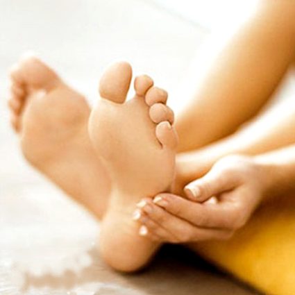
If you do not deal with the treatment yourself, after the regeneration of the bone tissue, protruding sections of the bone will not appear on the legs again, requiring surgical intervention. For prevention possible relapse disease shows a general massage and foot massage of medium intensity.
ethnoscience

At home, it is correct to treat osteochondropathy in a baby, without a preliminary examination and diagnosis in medical institution, almost impossible. Therefore, the planning of methods and techniques should be entrusted to the doctor. This will not only restore health to the legs, but also avoid similar problems later.
It occurs much more often than in adults. At risk are girls seven to eight years old and boys nine to eleven. Professional athletes and adults who are actively involved in sports are susceptible to pathology.
The main reason for the development of Schinz's disease is malnutrition of bone tissues and aseptic neurosis. Doctors associate secondary manifestations with the resorption of individual sections of the bones and their subsequent replacement. Osteochondropathies account for 2.7% of orthopedic pathologies. Schinz's disease was first described by the Swedish surgeon Haglund at the beginning of the last century.
So far, doctors do not have a common opinion on why osteochondropathy of the calcaneus occurs, but common factors can be distinguished. Among them:
- improper functioning of the endocrine glands;
- metabolic disorders (especially metabolic processes indispensable for normal operation body substances);
- poor absorption of calcium;
- trauma;
- increased physical activity.
Although it most often occurs in children, it can also affect adults. Especially if they are actively involved in sports (and are prone to injury) or have certain health problems (the bones do not absorb calcium well, nutrient metabolism is disturbed, and so on).
Symptoms
Osteochondropathy of the calcaneal tuber can develop in different ways - in some people the disease immediately becomes acute, in others long time can proceed sluggishly, almost asymptomatically. acute form characterized by severe pain, which is localized in the heel area and intensifies after physical exertion.
Other possible symptoms:

- swelling in the affected area;
- problems with flexion and extension of the foot;
- soreness of the affected area on palpation;
- , redness;
- limping when walking, sometimes it is difficult for a patient to stand on a sore leg without leaning on a cane, table or chair arm;
- pain at the point of attachment of the Achilles tendon to the heel bone;
- subsidence of pain in horizontal position(if the symptoms described above are in daytime, and at night during sleep subside or disappear altogether - we are talking about Shinz's disease)
Atrophy, hyperesthesia of the skin in the heel region, atrophy of the calf muscles are rare, but this possibility cannot be completely ruled out. Symptoms persist for a long time, in children they may disappear after the completion of the growth process.
How is the disease diagnosed?
 To diagnose osteochondropathy, an x-ray is taken. In the picture, there are violations of the structural patterns of the apophysis, fragmentation, distorted distances between the heel bone and the apophysis, the views are clear. On a sore leg, the unevenness of the contours will be more pronounced than on a healthy one. Before referring the patient to an x-ray, the doctor examines the legs and listens to complaints.
At severe forms Shinz's disease (calcaneal) x-ray the separation of parts will be clearly expressed marginal bone. Also, this pathology is always accompanied by an increase in the distance between the apophysis and the heel bone.
In some cases, the doctor prescribes differential diagnosis. Its passage will allow to exclude the presence of other pathologies with similar symptoms and similar changes in the bone.
Treatment
Treatment of osteochondropathy of the calcaneus in children and adults, the doctor prescribes after examination, taking into account individual features clinical picture- the complexity of the pathology, the patient's condition. AT acute stages complete rest of the foot that is affected is shown.
The main methods of treatment of Shinz disease (heel bone):
- Conservative - the load on the bone is reduced due to the use of a special splint with stirrups. If you are used to walking in flat shoes, you will need to replace them with shoes or shoes with a small (but not high!) Heel, but it is better to buy an orthopedic pair.
- Physiotherapy is ultrasound, electrophoresis.
- Warm compresses - they are convenient to use at home.
- The use of anti-inflammatory and analgesic ointments.
- Warm baths.
- Ozokerite applications.
And remember that the doctor should prescribe the treatment for you - only in this case it will be effective and give the desired results.
Shinz's disease is a pathological process associated with osteopathy (impaired ossification) of the apophysis (tubercle) of the heel bone. In medicine, this pathology is called "calcaneal osteochondropathy", because it causes destruction of the elements of the heel joint of the leg, caused by aseptic necrosis. Because of aseptic necrosis blood circulation of the cancellous bone of the heel is disturbed, which leads to destruction. This pathology is named Haglund-Shinz disease in honor of the Swiss orthopedic surgeon Petrik Haglund, who described in patients in 1907 a postero-superior seal in the heel area, and the scientist Schinz, who studied and described in more detail this pathology. Both adults and children can get sick.
AT childhood Schinz's disease is much more common among boys 9 to 11 years of age and among girls 7 to 8 years of age. Among adults, professional athletes and people with an active lifestyle are more susceptible to the disease. As a rule, the disease is benign in nature and does not impair the functionality of the joint during treatment. If treatment is not carried out, deforming arthrosis remains ( chronic pathology joint, with partial loss of mobility due to the altered shape of the bones).
The course of the disease
The heel bone is the largest spongy bone foot, an elongated and laterally flattened form, consisting of a diaphysis (body) and a protruding, easily palpable bone process (tubercle, or apophysis), affected by Shinz's disease. The calcaneus, due to its size, takes on almost the entire load experienced by the foot when a person walks, runs and jumps. In addition, the heel bone is an integral part of the joints of the foot and the place where several ligaments are attached (the long plantar ligament is attached to the bottom of the heel) and tendons (the Achilles tendon is attached to the heel apophysis). Due to the functions performed, the health of the heel bone is very important.
Despite the fact that the disease was discovered a long time ago, the mechanism of the course of the pathology and the root cause remain not fully elucidated to this day. There are only assumptions. It is believed that Schinz's disease begins with a violation of the blood supply to the tissues of the heel joint, as a result of which the adjacent tissues receive less nutrients and oxygen. Then the gradual destruction of the calcaneal tuber begins. The entire pathogenesis of Shinz's disease is accompanied by the following stages:
- 1. Aseptic necrosis. This is the necrosis of bone and joint tissues due to impaired circulation, and not due to infection in the blood.
- 2. Impression (depressed) fracture. On the this stage dead tissue disease becomes much larger, they lose their functions. As a result, the bone does not withstand the previous loads and some of its sections are pressed through, and then wedged into healthy areas of the bone.
- 3. Fragmentation - the division of the affected part of the bone into fragments.
- 4. Resorption of dead tissue.
- 5. Restoration (repair) of affected tissues. At this stage, they are replaced by connective scar tissue, instead of which a new bone will form over time.
Osteochondropathy is a disease from the group of degenerative-dystrophic pathologies, manifested by aseptic necrosis of certain groups of bones. More often than others, lesions of the tibia, apophyses of the vertebral body, femur and calcaneal bones occur. Osteochondropathy of the calcaneus in children is considered a common disease from this group and it affects mainly children aged 12-16 years, and if not treated seriously, then the pathology will bring many health problems in the future. In general, the feet due to increased load quite often they suffer from chondropathy, while the mechanism of pathology is based on the onset of aseptic necrosis of the metatarsal bone, calcaneal bones, tarsus, in addition, the scaphoid bones, the talus block can be affected.
The exact relationship with the origin of the disease has not yet been established. There are several theories of its origin. In childhood, there is often a connection between chondropathy and conditions when there are disturbances in the development of bone tissues. In adult patients, the relationship with increased loads on the musculoskeletal system is more characteristic. No exception is the situation when the patient fails to find out the causes of the origin of the disease. Let us consider in more detail such a topic as osteochondropathy of the calcaneus, its causes, symptoms of the disease and methods of treatment.
Classification
First, we will analyze what types of osteochondropathy are most common in order to understand the severity of this disease and the extent of damage to the structure of the musculoskeletal system. If we analyze the most common forms of pathology, we can distinguish Keller's disease, in which the head of the metatarsal bone or the navicular bone is affected. If the heel tuber area is involved in the pathological process, then Haglund-Shinz disease develops. Less common is a lesion in the region of sesamoid bones (a form of Renander-Muller disease).
Defeats occur spinal column, these include Scheuermann-Mau osteochondropathy, which is typical for young men under 18 years of age. A disease of the spinal column forms a curvature (kyphosis), which is manifested by pain in the back, a decrease in activity, and in adulthood - performance. When struck femur, then the pathology is called "Legg-Calve-Perthes disease", more often male children under 12 years of age suffer from it.

By the way, if we consider Keller's disease, then it affects not only the heads of the metatarsal bone, which is why there are two types of this form:
- Keller's disease type 1, when osteochondropathy occurs scaphoid, which is located in the center of the foot;
- Keller's disease type 2 is a form that affects the head of the metatarsal bone, located at the base of the toes.
Let us examine in detail the form in which the heel is affected - osteochondropathy of the calcaneal tuber.
Causes
As mentioned above, the exact causes of the formation of the disease are still being studied, but there are risk factors that provoke the onset of the disease. An important factor is an genetic predisposition when parents had osteochondropathy, and not necessarily the calcaneus. It could be osteochondropathy of the head of the metatarsal bone (Keller's disease type 2), navicular bone and other bones of the foot, lower extremities, spine.
In children, osteochondropathy of the calcaneal bones of the feet occurs in violations of the endocrine system, metabolic processes in the body, and hormonal disruptions. Often found congenital diseases, in which vitamins and trace elements are poorly absorbed, causing bones to suffer. In particular, the indigestibility of calcium affects the health of bones and joints, and since the foot takes over heavy load, especially the foot, then it is affected more often than other parts of the body.

Osteochondropathy (abbreviated dropopathy) also suffers from an increase in the load on the legs, in particular, on the feet. During excessive loads, muscles contract strongly, microtrauma occurs, the protection of bones and joints weakens, which increases the likelihood of inflammation. Since spongy bones suffer from osteochondropathy, the loads affect the narrowing of small vessels in the spongy bones. Most of the load increase occurs in fat people patients with impaired metabolism.
The last group of causes that cause osteochondropathy is trauma. It so happened that the feet are prone to injury, and most often it is squeezing during a fall from a height, due to an accident or injury at work.
Symptoms
It is worth noting that the symptoms of osteochondropathy affecting the calcaneus are found in girls, as they are more prone to hormonal disruptions. The main symptom of the disease is severe pain, as you know, the load on the foot goes to the heel and front section, fingers. Therefore, the appearance of symptoms of pain significantly worsens the quality of life, as the gait is disturbed, and rapid fatigue of the legs prevails.

The disease tends to show symptoms of soreness after exertion, that is, after walking, running or standing still for a long time. If both heels are affected, the child tries not to lean on them and walks on his toes. Naturally, this increases the load on the forefoot and, if not treated, the patient is at risk of developing flat feet, hallux valgus deformity of the 1st toe and curvature of 2-3 toes.
For children suffering from osteochondropathy of the bones of the foot, restrictions are introduced on physical activity, that is, you can not actively engage in sports. Lack of exercise leads to the process of atrophy. skin, muscles, sensitivity is disturbed. There are frequent cases when osteochondropathy of the heads of the metatarsal bones is combined with a lesion of the calcaneus, then patients cannot walk normally due to pain. Heads of 2-3 metatarsal bones, thumb are often affected.
Due to the fact that the nature of the gait is disturbed, not only the joints and muscles of the foot can suffer, with the progression of the disease, pain can also occur in the ankle, femoral, calf muscles and even in the region of the spine. If flat feet begin, then the chance of damage to the metatarsal bones increases, especially the 1-2 and 3rd.

The disease has several stages of development. Pathological process begins with aseptic necrosis, after which a state of impression fracture and fragmentation may develop. The next stage is based on the resorption of necrotic areas of the bone. In the future, repair, inflammation occurs, and if there is no treatment, then osteoarthritis develops, which will constantly progress.
Diagnostics
Diagnosis of osteochondropathy is based on the study of the totality clinical manifestations with results instrumental research. An X-ray examination is mandatory, and it is necessary to study not only the area of the calcaneus, but also other parts of the foot in order to exclude complications and comorbidities. With the help of x-ray diagnostics, arthrosis, traumatic bone lesions, osteochondropathy of the metatarsal head and calcaneus, as well as other structural units, are detected.
Careful diagnosis is needed in order to start treatment in a timely manner, since the lack of therapy for 2-3 years can lead to the development of arthrosis, bursitis, periostitis and other complications. In general, if therapy is carried out in a timely manner, then the chance of a complete cure is high.
Treatment
In most cases, osteochondropathy can be cured in a conservative way, that is, with the help of medicines, physiotherapy methods and physiotherapy exercises. The second treatment option is surgery. It is used in neglected forms or when ineffective conservative treatment. Of the minuses of the operation, there is a loss in the heel area of \u200b\u200bsensitivity in the future.
We will analyze how to treat the pathology conservatively. In order to facilitate treatment and reduce the manifestation of pain in the patient, orthopedic shoes or insoles are prescribed, which also prevent the development of flat feet. Among modern insoles, Solapro Viva can be distinguished. Viva insoles redistribute the load from the heel to the entire foot, while fixing the arches, preventing them from flattening. In addition, orthopedic insoles improve the process of blood supply in the foot area, reduce foot fatigue.
Treatment of osteochondropathy with symptoms of pain includes the use of NSAIDs. Patients are recommended to undergo treatment with one of the following drugs: Ibuprofen, Nimesulide.

Since children are more affected by this disease, the dosage, choice of drug and duration of therapy are determined individually by the doctor. At severe pain blockade with Novocain or Lidocaine in the heel area may be required. Also, with severe pain, it may be necessary to immobilize the heels with an orthosis, a plaster cast.
Necessarily treatment includes a course of physiotherapy. Electrophoresis procedures are prescribed for Novocaine and Analgin, ozokerite treatment. Effective compresses, baths with salt, soda and medicinal herbs. For successful therapy, it is recommended to take vitamins, especially from group B, calcium. AT recovery period you need to take a course of physiotherapy exercises, massage, to restore mobility to the feet.
Prevention and complications
Late treatment of osteochondropathy not only translates the disease into chronic form, but also threatens with other degenerative-dystrophic pathologies that affect the joints. Also, flat feet develop quite quickly, which remains for life, even after the treatment of osteochondropathy.

The treatment of advanced forms also leaves its mark. Necrotic lesion affects the sensitivity of the skin, therefore, after the course of treatment, these violations remain. The shape of the calcaneal tuber also changes, which can bulge, therefore, problems begin with the selection of shoes, walking.
In order to avoid osteochondropathy in children and adolescents, it is recommended to undergo medical examinations, especially with the appearance of pain in the feet, namely in the area of the calcaneus. For prevention with hereditary predisposition, it is necessary to perform a massage course (2-3 times a year), engage in physical therapy(regularly).
Shoes play an important role. Shoes should be comfortable, fit in size, not squeeze the feet. It is not recommended to wear shoes with very high heels, especially for girls in the period of growing up, that is, 14-17 years old. AT adolescence do not forget about vitamin therapy. Sufficient intake of vitamins B, D, calcium will strengthen the structure of bones.